Dura mater (Latin: dura mater)
Also called the pachymeninx
- Overview
- Cranial dura mater
- Spinal dura mater
- Reference websites
- (Wikipedia, websites with illustrations)
- Where it is found
- Related terms
Overview
What is the dura mater?
- Dura mater : the outermost and toughest; a dense fibrous membrane
- Arachnoid mater : a thin membrane between the dura and the pia mater; it has no blood vessels
- Pia mater : the innermost; a very thin membrane that follows every fold of the brain and spinal cord
| Space | Where it lies | What it is |
|---|---|---|
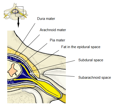
| Epidural space | Outside the dura mater | In the vertebral canal, a real space between the spinal dura and the wall of the canal |
| Subdural space | Between the dura and the arachnoid | Normally only a potential space; it opens up only in disease or injury (for example a subdural haematoma) |
| Subarachnoid space | Between the arachnoid and the pia mater | Filled with cerebrospinal fluid; it widens into cisterns in places |
Cranial dura mater
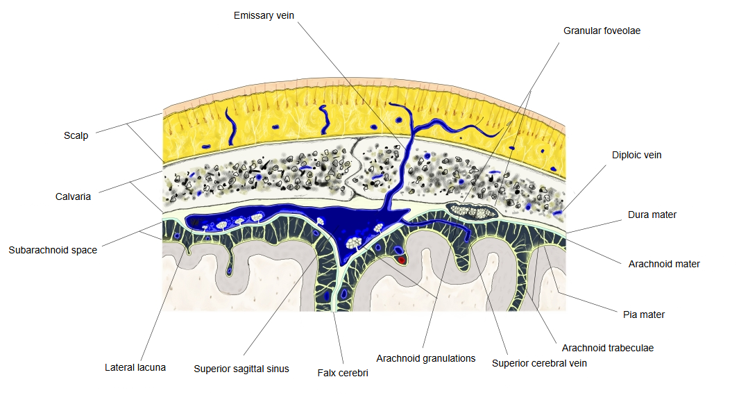
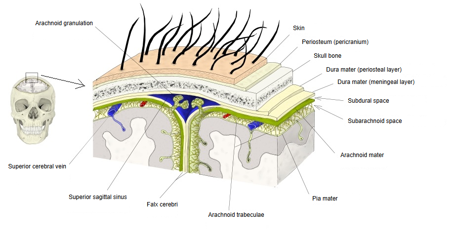
The cranial dura mater has an outer periosteal layer and an inner meningeal layer, fused into one sheet; unlike the spinal dura, it is not separated from the bone by a real space.
· It splits into its two layers only at the dural venous sinuses, the trigeminal cave and the endolymphatic sac.
· It is both the covering of the brain and the inner periosteum of the skull.
· Children: firmly attached to the skull. Adults: still firmly attached at the sutures and the cranial base; elsewhere only a potential (epidural) space lies between it and the bone.
The cranial dura mater is a single sheet of connective tissue formed by the fusion of an outer layer, the periosteum of the skull, and an inner meningeal layer. Only around the venous sinuses (such as the superior sagittal sinus), the trigeminal cave and the endolymphatic sac do the two layers separate.
It is both the covering of the brain and the inner periosteum of the skull
The cranial dura is therefore at the same time the covering of the brain and the periosteum lining the inside of the skull bones (the endocranium).
Children: firmly attached to the skull Adults: attached firmly at the sutures and cranial base; elsewhere only a potential space (epidural space) between it and the bone
In children the cranial dura is firmly attached to the skull. In adults it stays firmly attached at the cranial base and along the sutures between the bones of the skull; elsewhere it can be separated from the bone, but in health there is only a potential space, the cranial epidural space, which opens when blood collects there (an epidural haematoma). Between the dura and the bone run the vessels that supply the dura, mainly the middle meningeal artery and the middle meningeal veins. The dura is sensitive to pain; its sensory nerves come mainly from branches of the trigeminal nerve.
The cranial dura also forms sheaths and partitions.
It forms sleeves around the cranial nerves where they pass through the skull. Inside the cranial cavity its meningeal layer folds inwards to form partitions: the falx cerebri between the left and right cerebral hemispheres, the tentorium cerebelli between the cerebrum and the cerebellum, the falx cerebelli between the two cerebellar hemispheres and the diaphragma sellae over the pituitary gland.
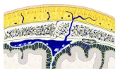 Meninges of the brain
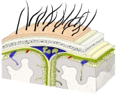 Layers from scalp to brain
Spinal dura mater
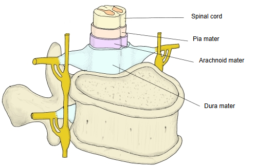
The spinal dura mater is a single tube (the dural sac); it does not line the vertebral canal
- Periosteum of the vertebral canal : lines the bone of the vertebral canal (some texts call it the outer layer of the spinal dura)
- Spinal dura mater (dural sac) : a tube that encloses the spinal cord and the roots of the spinal nerves; the dura mater proper
Above: attached to the margin of the foramen magnum, where it continues as the cranial dura mater.
Below: the sac encloses the spinal cord and the cauda equina and ends at about the level of the second sacral vertebra (sources give S1 to S3); a thin covering continues on the filum terminale and attaches to the back of the coccyx.
Sides: it extends along each spinal nerve root as a sleeve that blends with the epineurium of the spinal nerves.
The space between the periosteum and the dura mater is the epidural space; it contains fat, loose connective tissue and the internal vertebral venous plexus
The epidural space is also called the extradural space.
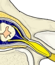 Meninges of the spinal cord
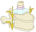 Spinal cord and its coverings
Reference websites
[Wikipedia]
· Dura mater, Wikipedia.
· Meninges, Wikipedia.
[Websites with illustrations]
· Human brain with the dura mater (photo) (Wikimedia Commons)

· Cranial dura mater (photo) (Wikimedia Commons)

· Spinal dura mater I (photo) (Wikimedia Commons)

· Spinal dura mater II (photo) (Wikimedia Commons)

Where it is found
Related terms
- Falx cerebri
- Granular foveolae
- Dural venous sinuses
- Superior sagittal sinus
- Middle meningeal veins
- Spinal cord
- Brain
References and sources
- Further reading: Dura mater, Wikipedia.
- Further reading: Meninges, Wikipedia.
- Federative International Programme for Anatomical Terminology (FIPAT). Terminologia Anatomica, 2nd ed. (TA2), 2019.