Pharyngeal constrictors
Also called Constrictors of pharynx or the pharyngeal constrictor muscles
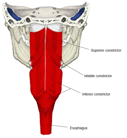
The muscle coat of the pharynx is divided into a longitudinal layer and a circular layer; the circular layer is made up of three parts: the superior, middle and inferior constrictors.
of the pharynx
- Circular muscle : narrows the pharynx: the pharyngeal constrictors.
- Longitudinal muscle : raises the pharynx: the pharyngeal elevators.
constrictors
(circular muscle)
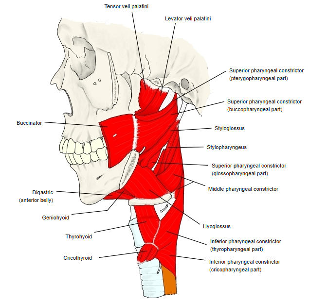
- Superior constrictor : the uppermost. From the pterygoid hamulus, the pterygomandibular raphe, the end of the mylohyoid line of the mandible and the side of the tongue. Above it, the wall is closed only by the pharyngobasilar fascia.
- Middle constrictor : in the middle. Fan-shaped; from the stylohyoid ligament and the lesser and greater horns of the hyoid bone.
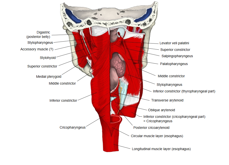
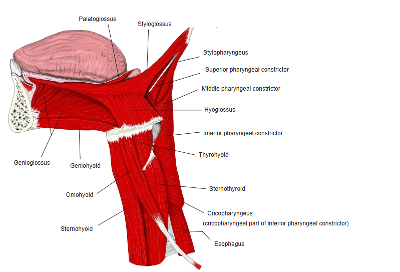
- Inferior constrictor : the lowest and thickest, in two parts: a thyropharyngeal part from the oblique line of the thyroid cartilage, and a cricopharyngeal part (cricopharyngeus) from the side of the cricoid cartilage, which acts as the upper esophageal sphincter.
· Stylopharyngeus and the glossopharyngeal nerve enter the pharynx through the gap between the superior and middle constrictors.
· The weak area between the thyropharyngeal and cricopharyngeal parts of the inferior constrictor (Killian's triangle) is where a pharyngeal pouch (Zenker's diverticulum) can form.
· The pharyngeal constrictors receive nerve fibres from the vagus nerve through the pharyngeal plexus; the inferior constrictor also receives branches of the external and recurrent laryngeal nerves.
- Formed by : the pharyngeal branches of the glossopharyngeal nerve, the pharyngeal branches of the vagus nerve, and the laryngopharyngeal branches of the superior cervical ganglion (sympathetic; one from the sympathetic trunk).
- Supplies
- 1. Muscles of the pharynx : all except stylopharyngeus (supplied by its own branch of the glossopharyngeal nerve): the superior, middle and inferior constrictors, palatopharyngeus and salpingopharyngeus.
- 2. Muscles of the palate : all except tensor veli palatini (supplied by the mandibular nerve through the nerve to medial pterygoid): levator veli palatini, musculus uvulae, palatoglossus and palatopharyngeus (which belongs to both groups).
They are circular muscles. All of them attach to the pharyngeal raphe in the midline of the back wall of the pharynx, where they meet their partners of the other side. They overlap like roof tiles from below upwards (the inferior overlaps the middle from outside, and the middle overlaps the superior) and narrow the cavity of the pharynx. During swallowing they contract one after another from above down and push the food into the esophagus.
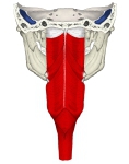 Pharyngeal constrictors (posterior)
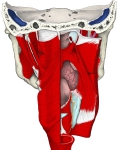 Muscles of pharynx (posterior)
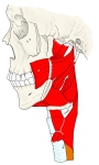 Pharyngeal constrictors (lateral)
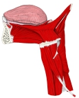 Tongue, pharynx and larynx (lateral)
Related terms
- Inferior pharyngeal constrictor
- Cricopharyngeus
- Stylopharyngeus
- Palatopharyngeus
- Pharyngeal branches of vagus nerve
- Pharynx
- Esophagus
References and sources
- Further reading: Superior pharyngeal constrictor muscle, Wikipedia.
- Further reading: Middle pharyngeal constrictor muscle, Wikipedia.
- Further reading: Inferior pharyngeal constrictor muscle, Wikipedia.
- Federative International Programme for Anatomical Terminology (FIPAT). Terminologia Anatomica, 2nd ed. (TA2), 2019.