Lymphoid nodule (Latin: Nodulus lymphoideus)
Also called the lymph nodule or the lymphoid follicle
- Overview
- Solitary lymphoid nodules
- Aggregated lymphoid nodules
- Do lymphoid nodules belong to the lymphatic system?
- Reference websites
- (Wikipedia, websites with illustrations)
- Related terms
Overview
· The similarly named lymph node is a lymphoid organ that contains lymphoid nodules inside it.
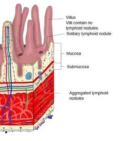
Solitary lymphoid nodules
· Its structure is like that of a lymph node, but it has no capsule; it also has no afferent lymphatic vessels and takes up antigens directly from the surface of the mucosa. Solitary nodules are scattered especially along the small and large intestine.
Cells other than B lymphocytes (this list may not be complete):
| Cell | Notes | |
|---|---|---|
| 1 | Follicular dendritic cells | Present antigens; maintain the interaction with B cells |
| 2 | Helper T cells | Help to activate B cells (especially the follicular helper T cells, Tfh) |
| 3 | Macrophages | Take up cells that have died by apoptosis; process antigens |
| 4 | Plasma cells | Produce antibodies (the mature stage) |
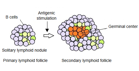
How much does a primary nodule grow after antigenic stimulation?
There is no fixed quantitative standard (such as "so many times larger") for how much a primary lymphoid nodule enlarges after antigenic stimulation, but histology and immunology give the following picture.
Size of primary and secondary lymphoid nodules (outline; rough values that vary with the site and state)
| Kind | State | Diameter (approx.) | Histological features | |
|---|---|---|---|---|
| 1 | Primary nodule | Resting (not yet stimulated by antigen) | about 100–200 µm | Stains evenly; no germinal centre; mainly naive B cells. |
| 2 | Secondary nodule | Activated by antigenic stimulation | about 300–800 µm (sometimes about 1 mm) | A clear germinal centre forms, surrounded by a darkly staining cap (mantle zone). |
Growth
After antigenic stimulation, naive B cells are activated and divide actively.
As a result, while the germinal centre forms, the diameter of the primary nodule is said to increase roughly two- to fivefold (based on histological observation).
Nodules may grow so large that neighbouring nodules appear to merge.
[ Points ]
· When a primary nodule becomes a secondary nodule, its form and function change.
· The enlargement is not mere swelling but the result of active division, selection and differentiation of B cells.
· It can grow to a size of several hundred micrometres up to almost 1 mm.
· This change is clearly visible under the microscope.
Aggregated lymphoid nodules
· Secondary aggregated lymphoid nodules
Primary aggregated lymphoid nodules are activated by antigenic stimulation, but usually the whole group is not activated at once;
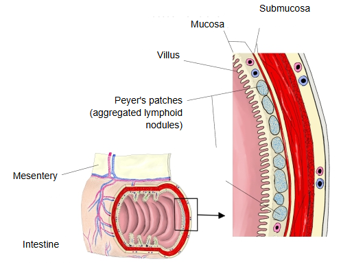
more often only some of the solitary nodules that make up the group are activated and form germinal centres. The best-known aggregated nodules are the Peyer's patches in the wall of the ileum, on the side opposite the mesentery.
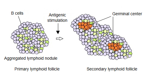
 Lymphoid nodules in the intestinal wall
 Peyer's patches
Do lymphoid nodules belong to the lymphatic system?
In short:
Lymphoid nodules belong to the lymphoid tissue, but not to the network of lymphatic channels.
That is, a lymphoid nodule is part of the lymphoid tissue, but not a part of the lymph pathway in the way that lymph nodes and lymphatic vessels are.
Sorting out the terms
| Group | Main structures | Relation to the lymph flow | |
|---|---|---|---|
| 1 | Lymphatic channels | Lymphatic vessels, lymph nodes, lymphatic trunks, thoracic duct and others | The system of channels that carries lymph through the body |
| 2 | Lymphoid tissue | Lymphoid nodules, spleen, thymus, tonsils, bone marrow and others | Tissues where immune cells gather; not necessarily on the course of the lymph flow |
What a lymphoid nodule is
A lymphoid nodule is a local, follicle-like cluster of lymphocytes, mainly B lymphocytes.
It lies on its own in connective tissue such as the lamina propria of the mucosa.
It has no afferent lymphatic vessels, so lymph does not flow into it (antigens reach it directly from the surface), although lymph drains away from it.
By contrast, a lymph node...
is a structure into which lymphatic vessels bring lymph and out of which they carry it; it filters the lymph and keeps immune watch over it.
Inside it there are also the lymphoid nodules of the cortex, but these are structural units within the lymph node. ⇒ It is more accurate to say that lymphoid nodules are also found inside lymph nodes.
Summary
| Point | Lymphoid nodule | Lymph node | |
|---|---|---|---|
| 1 | Nature | A local cluster of lymphocytes | An organ through which lymph flows in and out |
| 2 | Main function | Local immune responses (especially of B cells) | Filtering the lymph and immune surveillance |
| 3 | Location | Lamina propria of the mucosa and similar sites (part of the mucosa-associated lymphoid tissue) | On the course of the lymphatic vessels (arranged in regional groups) |
| 4 | Lymphatic vessels | No afferent vessels; lymph only drains away | Afferent and efferent lymphatic vessels attached |
Conclusion
Lymphoid nodules are part of the lymphoid tissue, but not of the lymphatic channels (lymphatic vessels). In other words, they belong to the lymphatic system in the wide sense, but they are not stations on the lymph flow as lymph nodes are.
Reference websites
[Wikipedia]
· Mucosa-associated lymphoid tissue, Wikipedia.
· Peyer's patch, Wikipedia.
[Websites with illustrations]
· Solitary lymphoid nodules (illustration) (IMAIOS e-Anatomy)
· Aggregated lymphoid nodules of the ileum (illustration) (IMAIOS e-Anatomy)
Related terms
References and sources
- Further reading: Lymphoid follicle, Wikipedia.
- Further reading: Peyer's patch, Wikipedia.
- Federative International Programme for Anatomical Terminology (FIPAT). Terminologia Anatomica, 2nd ed. (TA2), 2019.