Maxillary surface of perpendicular plate (Latin: facies maxillaris laminae perpendicularis ossis palatini)
- Nasal surface / medial surface : forms the back part of the lateral wall of the nasal cavity; it carries two ridges (ethmoidal crest and conchal crest).
- Maxillary surface / lateral surface : joins the maxilla (palatomaxillary suture).
Structure: the maxillary surface is divided into a large front part that covers the body of the maxilla and a back part made of rough areas.
surface
- Front part: covers the body of the maxilla : covers the back of the medial surface of the body of the maxilla; part of it closes the maxillary hiatus.
- Back part: rough areas : two vertical rough areas, front and back, with a smooth strip between them that forms the greater palatine canal.
- Front : joins the medial surface of the maxilla.
- Middle : runs down from the sphenopalatine notch; its lower part is the greater palatine groove, which with the matching groove of the maxilla forms the greater palatine canal, opening on the palate at the greater palatine foramen. Its upper part forms the medial wall of the pterygopalatine fossa.
- Back : joins the pterygoid process of the sphenoid bone.
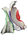 Maxillary surface and its contacts
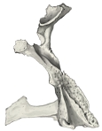 Right palatine bone (posterior view)
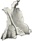 Palatine bone (maxillary surface)
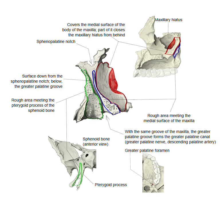
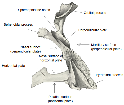
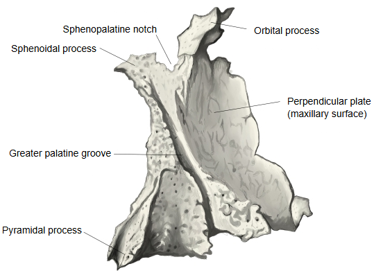
Behind the front part, which covers the back of the medial (nasal) surface of the body of the maxilla and closes part of the maxillary hiatus from behind and below (narrowing the opening of the maxillary sinus), the surface has two vertical rough areas: the front one meets the medial surface of the maxilla, the back one meets the pterygoid process of the sphenoid bone. Between them a smooth strip runs down from the sphenopalatine notch. Its upper part forms the medial wall of the pterygopalatine fossa; lower down it becomes the vertical greater palatine groove (see palatine grooves). With the matching groove on the maxilla this groove forms the greater palatine canal, which opens on the palate at the greater palatine foramen and carries the greater palatine nerve and the descending palatine artery. Small lesser palatine canals, usually two, branch from it, run through the base of the pyramidal process and open on its lower surface as the lesser palatine foramina.
Related terms
- Perpendicular plate
- Palatine bone
- Greater palatine canal
- Maxillary hiatus
- Pterygopalatine fossa
- Pterygoid process
- Pyramidal process
References and sources
- Further reading: Palatine bone, Wikipedia.
- Federative International Programme for Anatomical Terminology (FIPAT). Terminologia Anatomica, 2nd ed. (TA2), 2019.