Cortical labyrinth (Latin: labyrinthus corticis)
Also called the convoluted part of renal cortex
Overview
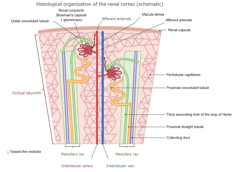
· The cortical labyrinth fills the spaces between the medullary rays; together they make up the renal cortex.
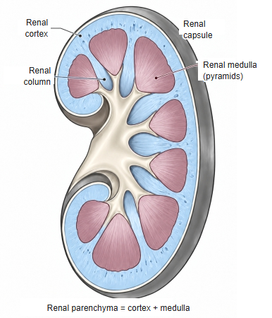
· The renal columns are parts of the renal cortex that reach down between the renal pyramids. Like the cortical labyrinth, they contain renal corpuscles and convoluted renal tubules.
· In microscope sections the round glomeruli identify the labyrinth and distinguish it from the medullary rays, which contain no renal corpuscles and appear as bands of straight tubules.
cortex
- Cortical labyrinth : renal corpuscles, proximal convoluted tubules, distal convoluted tubules, connecting tubules, juxtaglomerular apparatus, interlobular arteries and veins, afferent and efferent arterioles, capillariesabout 70–75%
- Medullary rays : proximal straight tubules, thick ascending limbs of the loop of Henle, collecting ducts, capillariesabout 25–30%
The percentages are approximate and differ between sources.
⇒ A more detailed diagram of the renal cortex
The diagram below gives a simple outline of the parts of the kidney.
- Renal cortex
- Renal corpuscle : glomerulus and Bowman's capsule
- Proximal tubule : reabsorbs water and nutrients from the primary urine (convoluted part)
- Distal tubule : adjusts salts and water (convoluted part)
- Cortical collecting duct : collects urine from several connecting tubules
- Renal medulla
- Renal pyramid : the cone-shaped tissue that forms the medulla; it contains:
- Loop of Henle : U-shaped part (proximal straight tubule, intermediate tubule, distal straight tubule)
- Medullary collecting duct : carries urine toward the renal papilla
- Papillary duct : opens at the tip of the renal papilla and empties urine into a minor calyx
- Renal pyramid : the cone-shaped tissue that forms the medulla; it contains:
Divisions
The table below compares the contents of the cortical labyrinth and the medullary rays.
| Component | Cortical labyrinth | Medullary ray |
|---|---|---|
| Location and appearance | Between the medullary rays. Renal corpuscles lie in it and the tubules are coiled. | Rays running from the medulla toward the capsule. Straight tubules run in bundles. |
| Nephron and collecting duct | ||
| Renal corpuscle (glomerulus + Bowman's capsule) | ○ All renal corpuscles lie here | – None |
| Proximal tubule | ○ Convoluted part | ○ Straight part (runs down into the medulla) |
| Distal tubule (straight part = thick ascending limb) | – None | ○ Rises from the medulla (thick ascending limb) |
| Distal tubule (convoluted part) | ○ Convoluted part; where it touches its own renal corpuscle it forms the macula densa | – None |
| Connecting tubule | ○ Joins the distal tubule to the collecting duct | – None |
| Collecting duct | Only its first part, where the connecting tubules join; it then turns into the medullary ray | ○ Cortical collecting duct (runs down into the medulla) |
| Blood vessels and other structures | ||
| Interlobular arteries and veins | ○ Run straight toward the capsule (between the lobules) | – None |
| Afferent and efferent arterioles | ○ Enter and leave the vascular pole of the renal corpuscle | – None |
| Juxtaglomerular apparatus | ○ Macula densa, juxtaglomerular cells, extraglomerular mesangial cells | – None |
| Capillaries | ○ Network of peritubular capillaries around the convoluted tubules | ○ Long, narrow capillary meshes along the straight tubules |
Note: One medullary ray with the cortical labyrinth around it forms one renal lobule.
Note: The medullary rays are bundles of straight tubules (straight parts and collecting ducts) that continue from the medulla into the cortex.
Reference websites
[Wikipedia]
· Renal cortex, Wikipedia.
· Nephron, Wikipedia.
[Websites with illustrations]
· Urinary system overview (Digital Histology)
· Plate 12.232 Kidney: Cortex (Atlas of Microscopic Anatomy)
· Tubules and collecting system (Digital Histology)
Related terms
- Medullary rays
- Renal sinus
- Renal parenchyma
- Kidney
- Interlobular arteries of kidney (cortical radiate arteries)
- Fibrous capsule of kidney
- Renal pelvis
References and sources
- Further reading: Renal cortex, Wikipedia.
- Further reading: Nephron, Wikipedia.
- Federative International Programme for Anatomical Terminology (FIPAT). Terminologia Anatomica, 2nd ed. (TA2), 2019.