Prechiasmatic sulcus (Latin: sulcus prechiasmaticus)
Also called: chiasmatic groove; sulcus chiasmaticus; optic groove
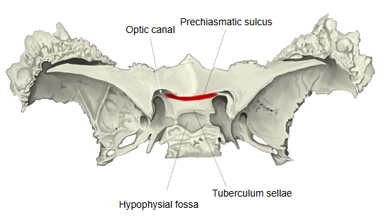
· A shallow groove runs across the upper surface of the body of the sphenoid in front of the tuberculum sellae; it is bounded in front by a low ridge at the back edge of the jugum sphenoidale. At each side it ends at the opening of the optic canal, which carries the optic nerve and the ophthalmic artery into the orbit. Despite its name, the optic chiasm usually lies above and behind the groove, over the diaphragma sellae, rather than in it. Its width and depth vary.
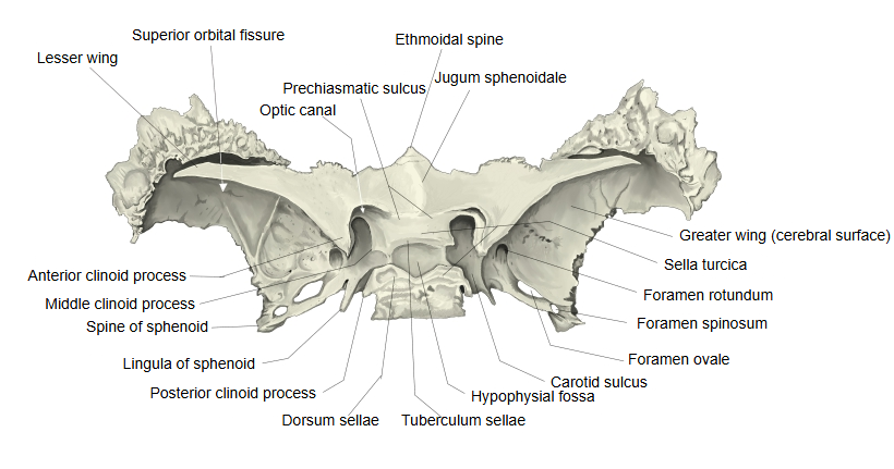
- Superior surface : faces the cranial cavity and forms the central part of the cranial fossae. Joins: the ethmoid bone in front; behind, the dorsum sellae forms the clivus with the basilar part of the occipital bone.Features: sella turcica, hypophysial fossa, tuberculum sellae, dorsum sellae, middle clinoid process, posterior clinoid process, prechiasmatic sulcus, carotid sulcus, sphenoidal lingula
- Inferior surface : forms the roof of the pharynx. Joins: the vomer (ala of vomer).
- Anterior surface : faces the nasal cavity. Joins: the ethmoid bone (and the orbital processes of the palatine bones).Features: sphenoidal crest (continuing below as the sphenoidal rostrum), sphenoidal concha (aperture of sphenoidal sinus)
- Posterior surface : a rough, four-sided surface. Joins: the occipital bone (basilar part).
 Sphenoid bone (superior)
 Sphenoid bone (superior), labelled
Reference websites
[Wikipedia]
· Chiasmatic groove, Wikipedia.
· Sphenoid bone, Wikipedia.
[Websites with illustrations]
· Chiasmatic groove (Wikimedia Commons)

· Sphenoid bone, upper surface (Gray's Anatomy) (Wikimedia Commons)

· The middle cranial fossa (TeachMeAnatomy)
Related terms
- Tuberculum sellae
- Optic canal
- Jugum sphenoidale
- Hypophysial fossa
- Sella turcica
- Body of sphenoid
- Sphenoid bone
References and sources
- Further reading: Chiasmatic groove, Wikipedia.
- Further reading: Sphenoid bone, Wikipedia.
- Federative International Programme for Anatomical Terminology (FIPAT). Terminologia Anatomica, 2nd ed. (TA2), 2019.