Latin: radius · one of the two bones of the forearm
Overview
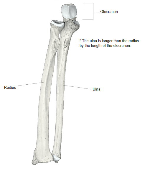
- The lateral bone of the forearm, on the thumb side; it lies parallel to the ulna.
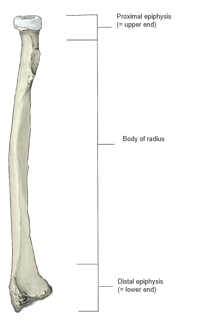
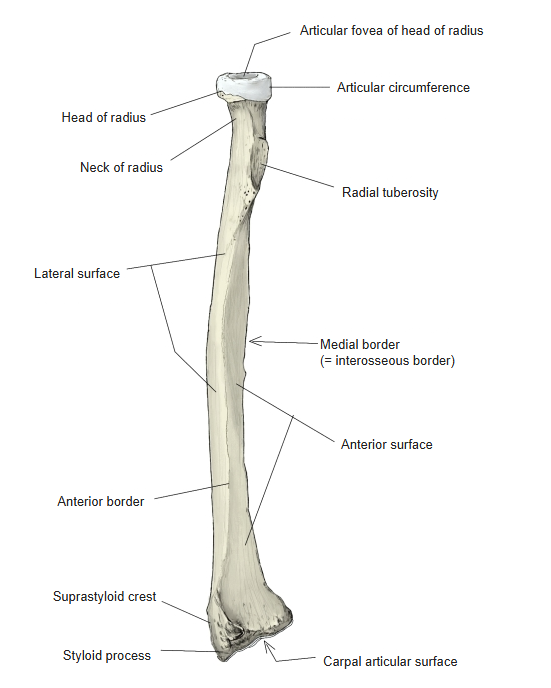
- Narrow at its upper end (head of radius) and broad at its lower end, which forms the greater part of the wrist joint.
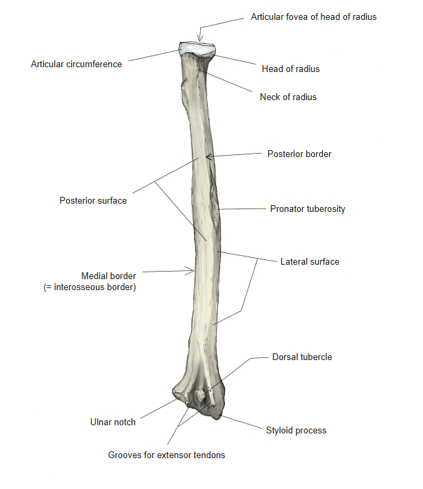
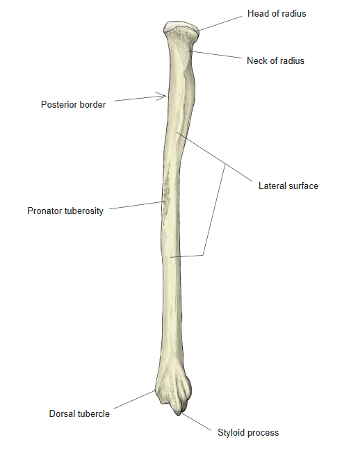
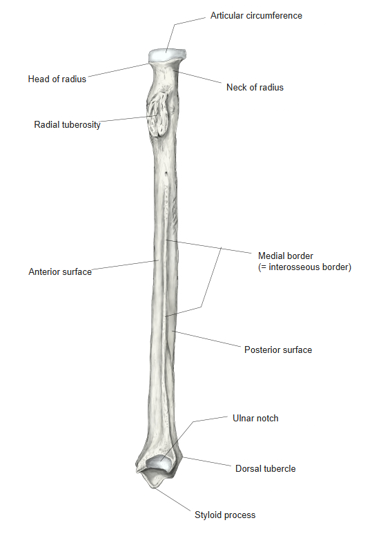
- Its shaft (body of radius) has three borders and three surfaces.
- Pronation and supination: the radius rotates around the ulna at the proximal and distal radioulnar joints. In pronation the two bones cross; in supination they lie parallel.
- Development: ossification centres appear in the shaft at about the 8th fetal week, in the distal epiphysis at about 2 years and in the proximal epiphysis at about 5 years; the proximal epiphysis fuses with the shaft at about 17–18 years and the distal epiphysis at about 20 years.
- Anomaly: the radius may be absent; the thumb is then usually absent as well.
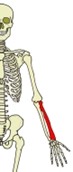
Bones of the left upper limb 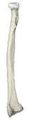
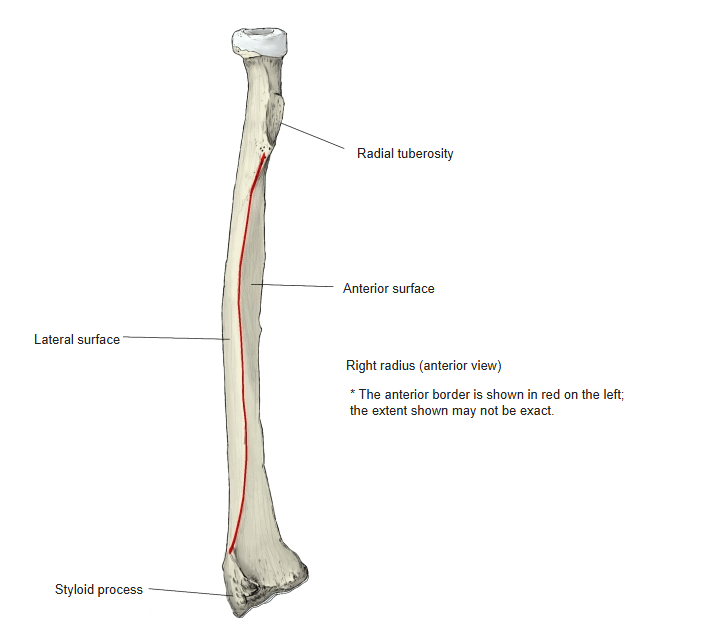
Right radius (anterior view)
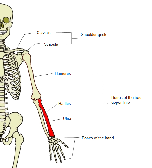
- Bones of the upper limb (one side)
- Shoulder girdle
- Clavicle
- Scapula
- Free upper limb
- Arm
- Humerus
- Forearm
- Radius ← thumb side
- Ulna ← little-finger side
- Hand
- Carpal bones (8)
- Metacarpal bones (5)
- Phalanges (14)
- Arm
- Shoulder girdle
Illustrations
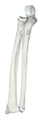
Right radius and ulna 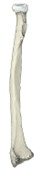
Right radius (anterior) 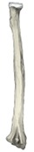
Right radius (posterior) 
Right radius (lateral) 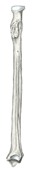
Right radius (medial) 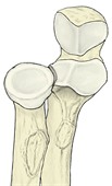
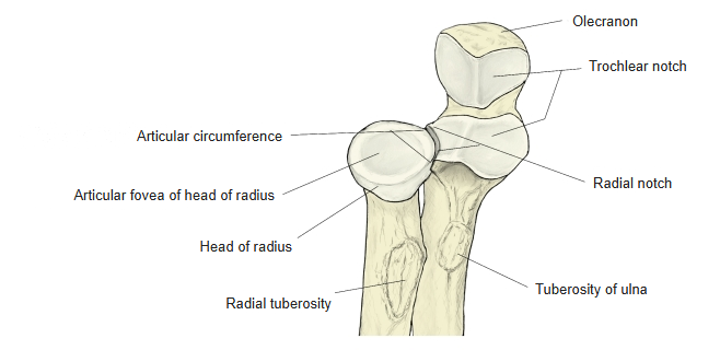
Radius and ulna (proximal ends) 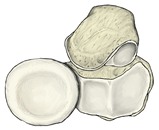
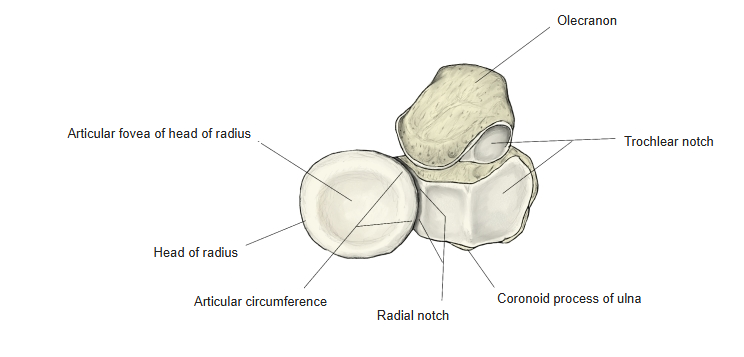
Radius and ulna, seen from above 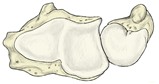
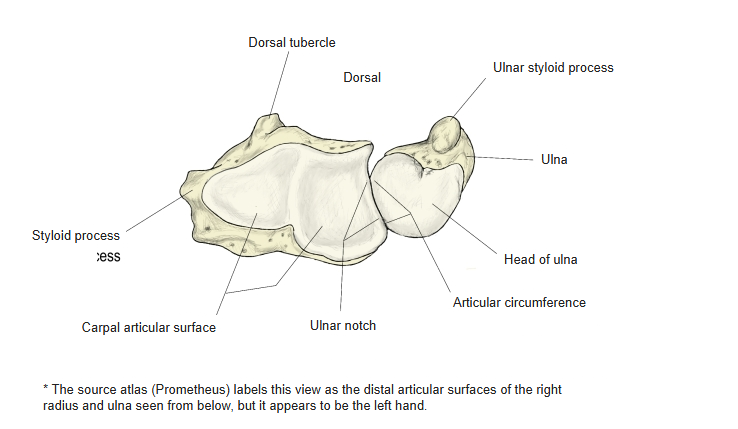
Radius and ulna, seen from below 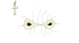
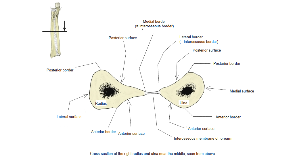
Cross-section of the forearm bones
Articulations
| # | Bone | Joint |
|---|---|---|
| 1 | Humerus | Humeroradial joint |
| 2 | Ulna | Proximal radioulnar joint |
| 3 | Ulna | Distal radioulnar joint |
| 4 | Scaphoid | Radiocarpal joint |
| 5 | Lunate | |
| 6 | Triquetrum |
Elbow joint
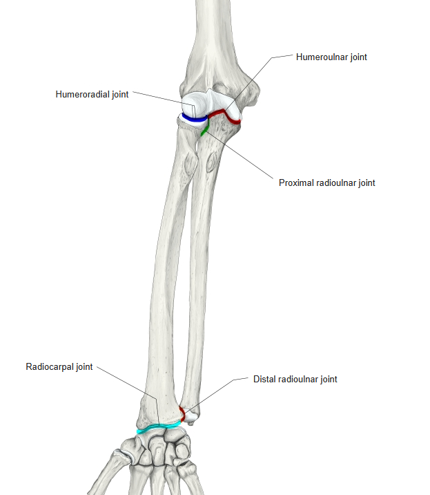
Humeroradial joint: articular fovea of the head ↔ capitulum of the humerus. Proximal radioulnar joint: articular circumference ↔ radial notch of the ulna. Distal radioulnar joint: ulnar notch ↔ articular circumference of the ulna. Radiocarpal joint: carpal articular surface of the radius and the articular disc ↔ scaphoid, lunate and triquetrum.
Ligaments
The following ligaments attach to the radius. The list is intended to be comprehensive but may omit minor ligaments.
| # | Ligament | Connects |
|---|---|---|
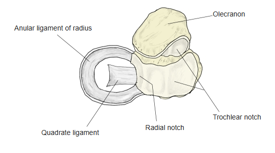
| 1 | Quadrate ligament | Neck of radius – radial notch of ulna (proximal radioulnar joint) |
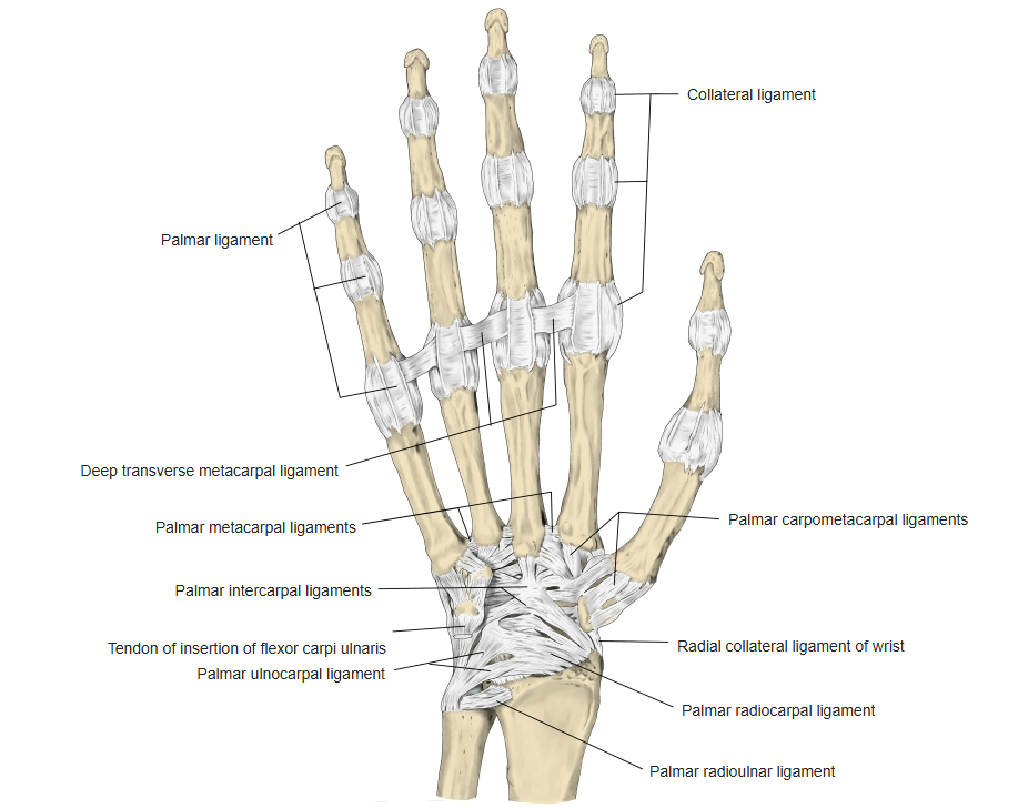
| 2 | Palmar radioulnar ligament | Ulnar notch of radius – head of ulna (distal radioulnar joint) |
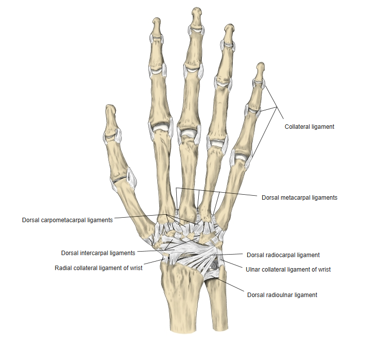
| 3 | Dorsal radioulnar ligament | Medial side of the lower end of radius – styloid process of ulna |
| 4 | Dorsal radiocarpal ligament | Styloid process of radius → scaphoid, lunate, triquetrum |
| 5 | Palmar radiocarpal ligament | Anterior margin of the lower end and styloid process of radius → scaphoid, lunate, triquetrum (some fibres reach the capitate) |
| 6 | Radial collateral ligament of the wrist | Styloid process of radius → scaphoid, trapezium |
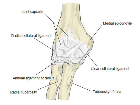
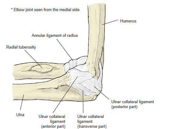
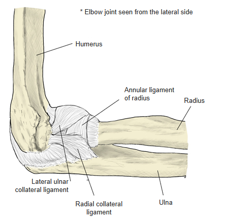
The radial collateral ligament of the elbow does not attach to the radius directly; it blends with the anular ligament of the radius.
Right elbow joint (posterior) 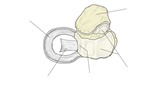
Quadrate ligament 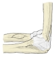
Right elbow joint (medial) 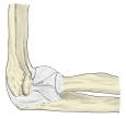
Right elbow joint (lateral) 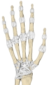
Right hand (palm) 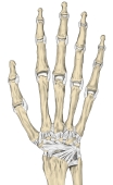
Right hand (dorsum)
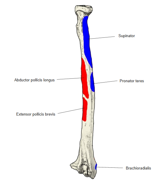
Muscle attachments
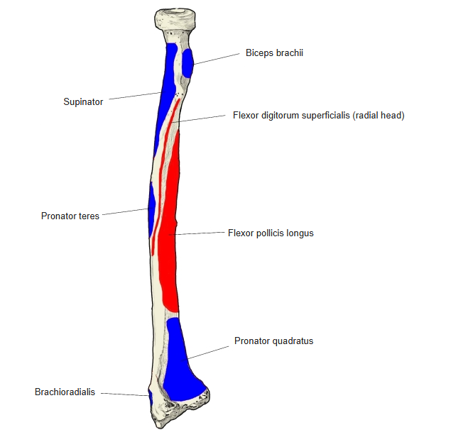
- Origin of: Abductor pollicis longus, Flexor digitorum superficialis, Extensor pollicis brevis, Flexor pollicis longus
- Insertion of: Biceps brachii, Brachioradialis, Pronator teres, Supinator, Pronator quadratus
Origins: abductor pollicis longus — posterior surface (middle); flexor digitorum superficialis (radial head) — anterior oblique line; extensor pollicis brevis — posterior surface (distal to the middle); flexor pollicis longus — anterior surface (around the middle). Insertions: biceps brachii — radial tuberosity; brachioradialis — above the styloid process; pronator teres — pronator tuberosity; supinator — lateral surface, upper third; pronator quadratus — anterior surface, lower quarter.

Muscle attachments (anterior) 
Muscle attachments (posterior)
Borders
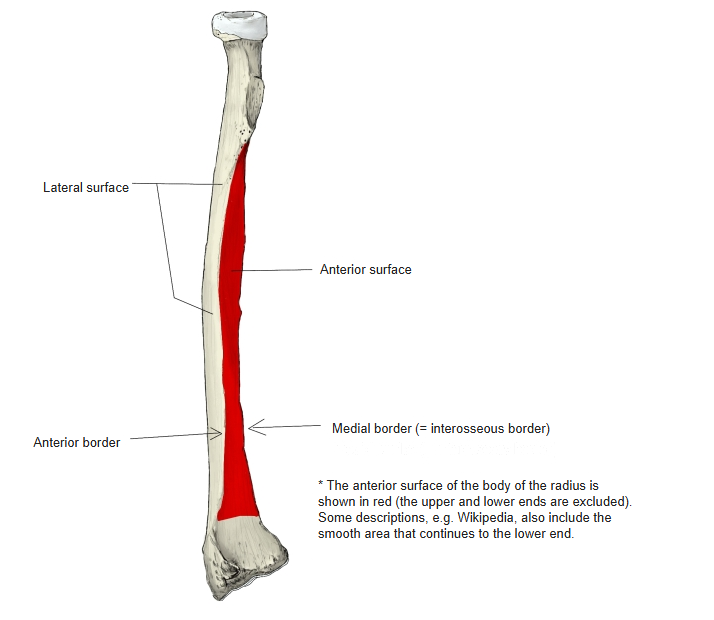
- Anterior border
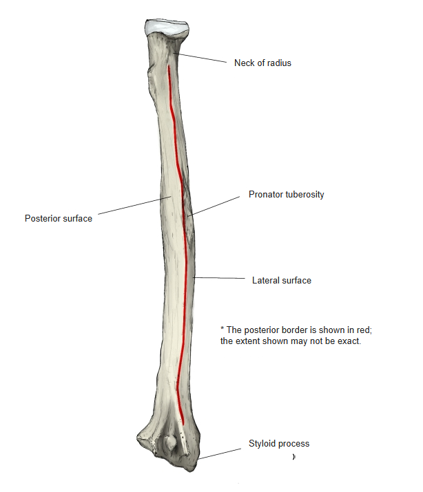
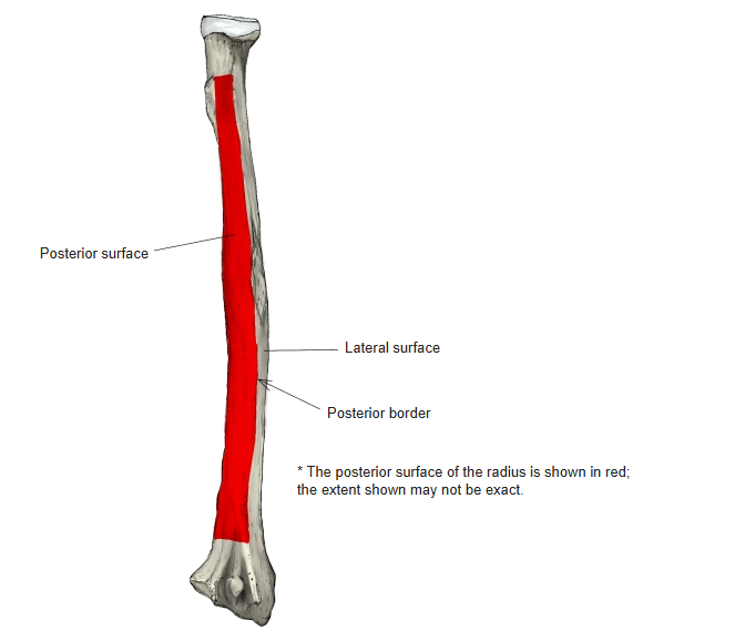
- Posterior border
- Medial border (interosseous border) — gives attachment to the interosseous membrane.
Borders (anterior view) 
Borders (posterior view)
Surfaces
- Anterior surface
- Posterior surface
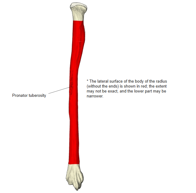
- Lateral surface
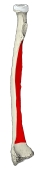
Anterior surface 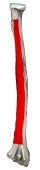
Posterior surface 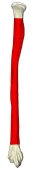
Lateral surface
References and sources
- Further reading: Radius (bone), Wikipedia.