Subperitoneal fascia
Also called Extraperitoneal fascia or the extraperitoneal connective tissue
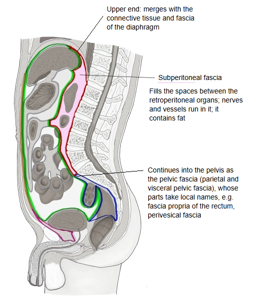
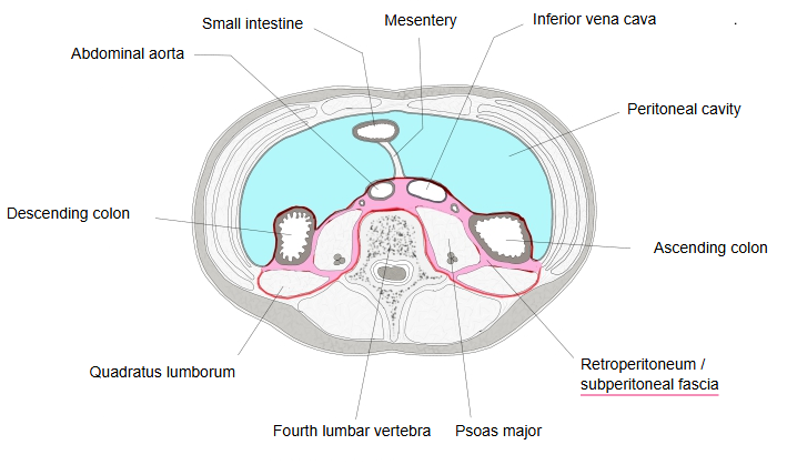
The subperitoneal fascia is the connective tissue that fills the spaces between the organs of the retroperitoneum; nerves and blood vessels run through it.
Retroperitoneum: the largest extraperitoneal space, running up and down behind the abdominal cavity and the peritoneal cavityIt lies between the parietal peritoneum and the walls of the abdomen and pelvis
· Although it is called a 'fascia', it is not the thin sheet that wraps round a muscle: nerves and blood vessels pass through it,
and it also contains fat, so it is a thick layer of loose connective tissue.
Function: it supports and protects the organs, nerves and blood vessels embedded in it. Sources differ in naming and limits: 'extraperitoneal fascia', 'subperitoneal fascia' and 'extraperitoneal tissue' are used for the whole layer or for parts of it.
Upper end: it passes over into, and merges with, the connective tissue and fascia of the diaphragm. Lower end: near the pelvic inlet it continues into the pelvic fascia.
Subperitoneal
fascia
fascia
- Pelvic fascia : continues from the subperitoneal fascia near the pelvic inlet
- Parietal pelvic fascia : the fascia that covers the pelvic walls (obturator internus, piriformis and others) + the presacral fascia.
- Visceral pelvic fascia : covers the surfaces of the pelvic organs and forms the connective tissue between them; nerves and blood vessels run in it. Its parts take names from the organ they surround, for example the fascia propria of the rectum, the vesical fascia and the prostatic fascia.
Related terms
- Extraperitoneal space
- Subperitoneal space
- Parietal pelvic fascia
- Transversalis fascia
- Parietal peritoneum
- Extraperitoneal organs
- Peritoneum
References and sources
- Further reading: Extraperitoneal fat, Wikipedia.
- Federative International Programme for Anatomical Terminology (FIPAT). Terminologia Anatomica, 2nd ed. (TA2), 2019.