Subperitoneal space (Latin: spatium subperitoneale)
Overview
The subperitoneal space is one of the parts of the extraperitoneal space; as its name says, it lies below the peritoneum (the peritoneal cavity) in the pelvis.
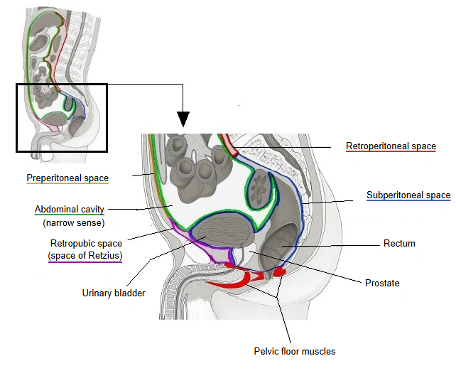
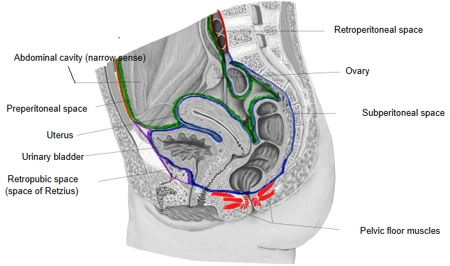
The layer of loose connective tissue just outside the peritoneal cavity (the abdominal cavity in the narrow sense)
- Retroperitoneal space : = retroperitoneum; the largest area, between the peritoneum of the back of the abdominal cavity and the posterior abdominal wall; it contains the kidneys, suprarenal glands, pancreas and abdominal aorta.
- Retropubic space : = space of Retzius; the area rich in fat at the front and lower part, between the pubis and the urinary bladder.
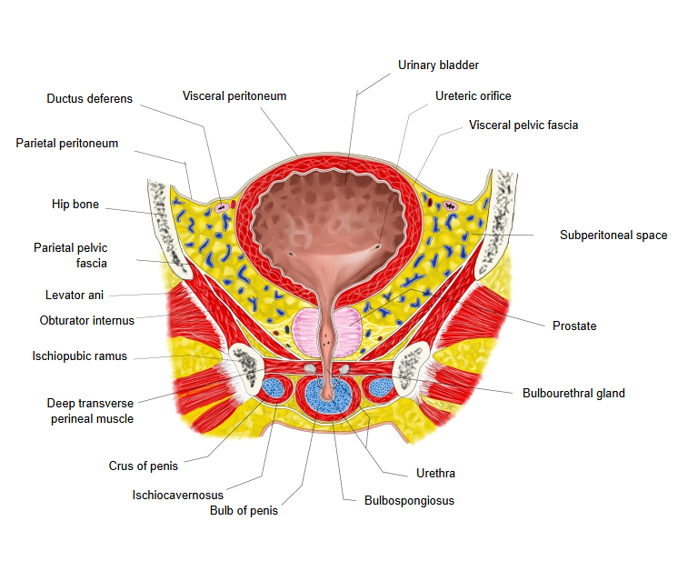
- Subperitoneal space : the area between the floor of the peritoneal cavity and the floor of the pelvis (pelvic floor muscles). Organs: rectum (middle and lower parts), urinary bladder (lower part), prostate, seminal glands, cervix of the uterus, part of the vagina.
- Preperitoneal space : the area at the front of the abdomen between the parietal peritoneum and the fascia of the abdominal wall.
Boundaries: above, the parietal peritoneum (the floor of the peritoneal cavity); below, the pelvic floor muscles (levator ani); at the sides, the pelvic walls (sacrum, hip bones and others).
It continues without a clear border into the retropubic space in front and the retroperitoneal space behind and above, and surrounds the organs that lie at the bottom of the pelvis. It is filled with loose connective tissue and fat.
Sources differ in where they draw the borders between the subperitoneal, retropubic and retroperitoneal spaces.
The subperitoneal space holds organs of the urinary, genital and digestive systems (the lower rectum and the anorectal junction).
List of organs
Organs that lie in the subperitoneal space are sometimes called "subperitoneal organs", but this is not an official anatomical term.
| Organ | Notes | |
|---|---|---|
| 1 | Rectum (middle and lower parts) | The upper part is covered by peritoneum in front and at the sides, the middle part only in front; the lower part has no peritoneal covering and lies entirely below the peritoneum. |
| 2 | Anal canal | Only its upper end (the anorectal junction) reaches the space; most of the canal lies below the pelvic floor, in the perineum. |
| 3 | Urinary bladder | Only its upper surface is covered by peritoneum; as a whole it is an extraperitoneal organ. |
| 4 | Ureter (pelvic part) | Continues from the retroperitoneal space into the subperitoneal space. |
| 5 | Prostate (male) | Just below the bladder; entirely an organ of the subperitoneal space. |
| 6 | Seminal gland | Behind the bladder and in front of the rectum, below the peritoneum. |
| 7 | Ampulla of the ductus deferens | The end part of the ductus deferens; it lies against the back of the bladder. |
| 8 | Cervix of the uterus (female) | The body of the uterus has a peritoneal covering, but the cervix lies below the peritoneum. |
| 9 | Vagina (most of it) | Only the upper part of its back wall is close to the peritoneum; it is almost entirely a subperitoneal organ. |
Nerves and vessels
| Structure | Notes | |
|---|---|---|
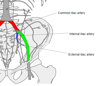
| 1 | Internal iliac artery | The main artery of the pelvic organs; one of the two terminal branches of the common iliac artery. |
| 2 | Pelvic venous plexuses | Well developed around the bladder, prostate and rectum. |
| 3 | Inferior hypogastric plexus | The autonomic nerve plexus of the pelvic organs. It is fed from above by the superior hypogastric plexus, which itself lies outside this space, in the retroperitoneal space in front of the sacral promontory. |
Reference websites
[Wikipedia]
· Pelvic cavity, Wikipedia.
[Websites with illustrations]
Related terms
- Rectum
- Urinary bladder
- Prostate
- Internal iliac artery
- Pelvic venous plexuses
- Inferior hypogastric plexus
- Parietal peritoneum
References and sources
- Further reading: Pelvic cavity, Wikipedia.
- Further reading: Extraperitoneal space, Wikipedia.
- Federative International Programme for Anatomical Terminology (FIPAT). Terminologia Anatomica, 2nd ed. (TA2), 2019.