Retroperitoneum (Latin: spatium retroperitoneale)
Also called: retroperitoneal space
- Overview
- Boundaries
- Retroperitoneal organs
- Vessels and nerves
- Relation to the adventitia
- Reference websites
- (Wikipedia, websites with illustrations)
- Related terms
Overview
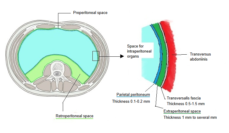
· Although its name contains "peritoneum", it is not a membrane but a region: it lies behind the parietal peritoneum and in front of the posterior abdominal wall. It holds the kidneys, suprarenal glands and ureters, parts of the gut, the abdominal aorta, the inferior vena cava, lymph nodes and nerves, all packed in fat and loose connective tissue.
peritoneal
space
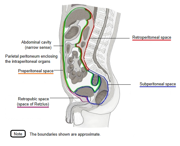
- Retroperitoneal space : = retroperitoneum; the largest area, behind the peritoneal cavity, between the parietal peritoneum in front and the posterior abdominal wall behind; it contains the kidneys, suprarenal glands, most of the pancreas, the abdominal aorta and the inferior vena cava.
- Retropubic space : = space of Retzius; the area rich in fat at the front and lower part, between the pubis and the urinary bladder; it lets the bladder expand.
- Subperitoneal space : the area between the floor of the peritoneal cavity and the floor of the pelvis (pelvic floor). Organs: rectum (lower part), urinary bladder (lower part), prostate, seminal glands, cervix of the uterus, part of the vagina.
- Preperitoneal space : the area at the front of the abdomen between the parietal peritoneum and the fascia of the abdominal muscles.
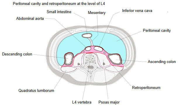
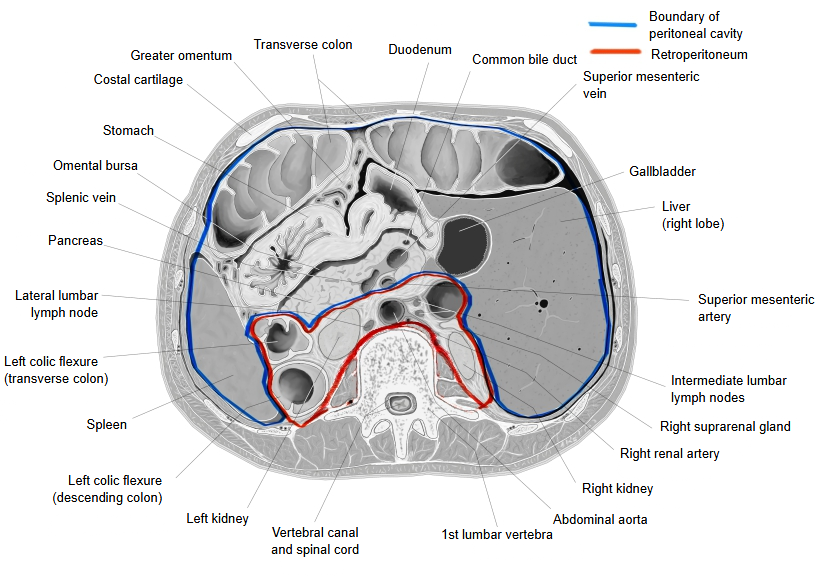
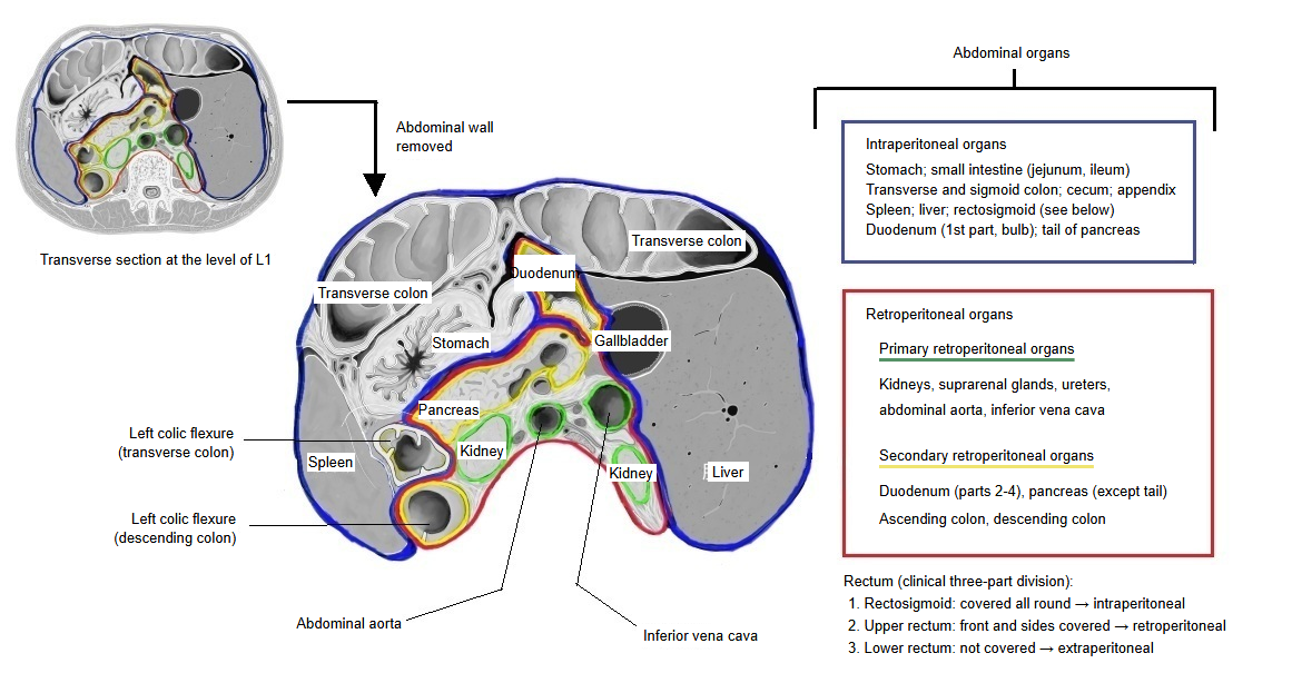
In the figure on the left below, the region of the retroperitoneum at the level of the fourth lumbar vertebra is shown in pink. The region of the abdominal cavity looks quite different at other levels, because the organs it holds change with the level.
Boundaries
The structures that bound the retroperitoneum are listed below.
| In front | Parietal peritoneum | The boundary with the peritoneal cavity (abdominal cavity) | |
| Behind | Posterior abdominal wall | Vertebrae | The front of the bodies of the lumbar vertebrae (mainly L1–L5) and of the intervertebral discs |
| Muscles | Psoas major, quadratus lumborum, iliacus | ||
| Fascia of transversus abdominis | Together with its aponeurosis | ||
| At the sides | Muscles and fascia | The muscles of the lateral abdominal wall (internal oblique, external oblique, transversus abdominis) | |
| Above | Diaphragm | Vessels and the oesophagus pass through openings (hiatuses) in the diaphragm, so it does not close the retroperitoneum completely. | |
| Below | Pelvic cavity | No boundary: it is continuous with the pelvis. | |
Retroperitoneal organs
peritoneal
organs
- Primary retroperitoneal organs : lie in the retroperitoneum from the start of development; their whole surface is covered by an adventitia and they are fixed to the posterior abdominal wall.
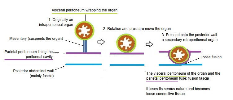
- Secondary retroperitoneal organs : lie within the peritoneum (abdominal cavity) at first ⇒ during development they press against the peritoneum and move into the retroperitoneum.
Primary retroperitoneal organs
The main features of the primary retroperitoneal organs are as follows.
· They lie in the retroperitoneal space from the start of development, and they differentiate and grow there.
· From the start they are surrounded by an adventitia, and through this adventitia they are fixed to the posterior abdominal wall (mainly to its fascia).
· Their front touches the parietal peritoneum. (This contact should be told apart from the fusion of the secondary retroperitoneal organs to the parietal peritoneum.)
The primary retroperitoneal organs are listed below; vessels and nerves are not included.
| Name | Mobility | Notes | |
|---|---|---|---|
| 1 | Kidney | large | Wrapped together with the suprarenal gland and fat in the renal fascia (Gerota's fascia); the two are separated by a septum. Loosely attached to the posterior abdominal wall by loose connective tissue. |
| 2 | Suprarenal gland | small | Closely related to the kidney, but it moves less than the kidney because it is firmly attached to the diaphragm. |
| 3 | Ureter | differs along its length | The upper part moves a lot with the kidney; lower down it tends to be more fixed. |
Vessels and nerves
| Vessels | 1 | Abdominal aorta | From the aortic hiatus of the diaphragm to its division into the common iliac arteries |
| 2 | Inferior vena cava | Formed by the union of the common iliac veins at the level of the fifth lumbar vertebra | |
| 3 | Renal arteries and veins | Run sideways to their organ from the start | |
| 4 | Suprarenal arteries and vein | ||
| 5 | Testicular / ovarian arteries and veins | Gonadal vessels | |
| 6 | Lumbar arteries and veins | Supply the muscles of the back | |
| Nerves | 1 | Lumbar plexus | Nerves that run within and on the surface of psoas major to the hip joint and the lower limb: the femoral nerve, the obturator nerve and others |
| 2 | Sympathetic trunk | Runs down each side of the vertebral column |
Secondary retroperitoneal organs
The main features of the secondary retroperitoneal organs are as follows.
· At first they are intraperitoneal organs with a mesentery; early in fetal life they move towards the back.
· Later in fetal life the organs have almost reached their final position.
· After that, the fixation of the organs, with their front covered by the parietal peritoneum, is almost completed.
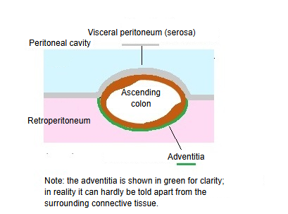
On the back of the organ, the peritoneum (serosa) that was there at first regresses and becomes an adventitia.
The secondary retroperitoneal organs are listed below.
| Name | Relation to the peritoneum | |
|---|---|---|
| 1 | Duodenum (parts 2–4) | The first part is covered by peritoneum and is sometimes counted as an intraperitoneal organ, but it has no mesentery, so it differs from a typical intraperitoneal organ. |
| 2 | Pancreas (head and body) | The tail is covered by peritoneum and is counted as an intraperitoneal organ. |
| 3 | Ascending colon | Its front and sides are covered by the parietal peritoneum. |
| 4 | Descending colon | Its front and sides are covered by the parietal peritoneum. |
| 5 | Rectum (upper and middle parts) | The front and sides of the upper part, and the front of the middle part, are covered by the parietal peritoneum. The lower part is a subperitoneal organ. |
· Sources differ on the caecum, which is usually described as intraperitoneal.
Relation to the adventitia
The table below shows simply how the retroperitoneal organs are related to the two coverings.
| Organ | Serosa | Adventitia |
|---|---|---|
| Kidneys, suprarenal glands, ureters | none | all round |
| Ascending and descending colon | front only | back only |
| Duodenum | part of the front | most of the surface |
| Pancreas | part of the front | most of the surface |
and the
peritoneum
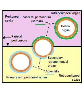
- Intraperitoneal organs : very mobile. Stomach, small intestine (jejunum, ileum), transverse colon, sigmoid colon, liver, spleen.
- Secondary retroperitoneal organs : first inside the peritoneal cavity ⇒ fused to the posterior wall during development. Duodenum (parts 2–4), pancreas (except the tail), ascending colon, descending colon.
- Primary retroperitoneal organs : firmly fixed. Kidneys, suprarenal glands, ureters, abdominal aorta, inferior vena cava.
Reference websites
[Wikipedia]
· Retroperitoneal space, Wikipedia.
· Peritoneum, Wikipedia.
[Websites with illustrations]
· Retroperitoneal space at the level of the kidneys (figure) (Journal of Clinical Imaging Science)
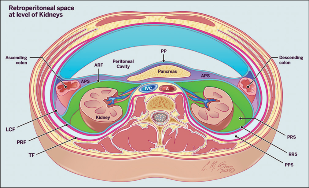
· Retroperitoneal space with kidneys and fascial layers (diagram) (Shutterstock)

· Retroperitoneal spaces and fasciae (figure) (Springer Nature)

Related terms
- Retroperitoneal organs
- Primary retroperitoneal organs
- Extraperitoneal space
- Intraperitoneal organs
- Parietal peritoneum
- Peritoneal cavity
- Tunica adventitia
References and sources
- Further reading: Retroperitoneal space, Wikipedia.
- Further reading: Peritoneum, Wikipedia.
- Federative International Programme for Anatomical Terminology (FIPAT). Terminologia Anatomica, 2nd ed. (TA2), 2019.