Latin: organum vestibulocochleare; auris
Overview
- The organ of hearing and balance. It turns sound waves and movements of the head into nerve signals.
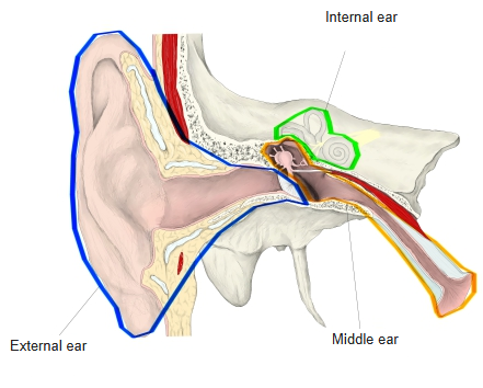
- Three parts: the external ear, the middle ear and the internal ear.
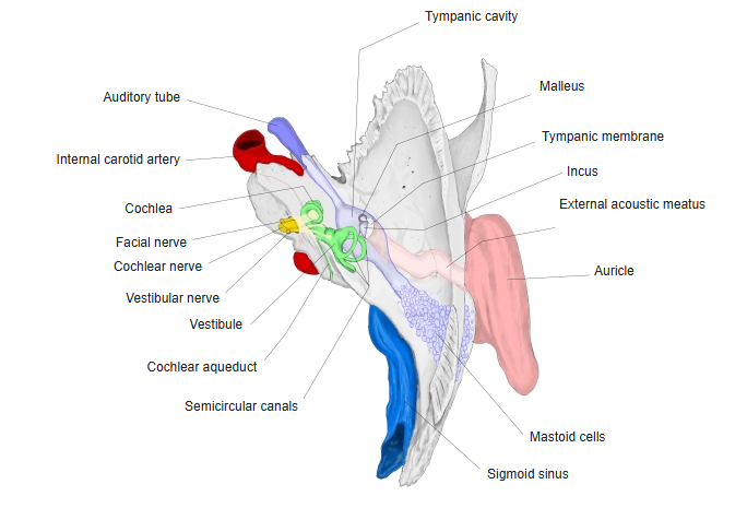
- Apart from the auricle, almost the whole ear lies inside the temporal bone; the internal ear is buried in its petrous part.
- The external and middle ear conduct sound; the internal ear contains the receptors for hearing and for balance.
- Signals travel to the brain in the vestibulocochlear nerve (cranial nerve VIII).
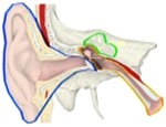
External, middle and internal ear 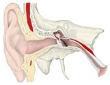
Ear, coronal section
Pages on the ear
Parts of the ear
| Part | Main structures | Role |
|---|---|---|
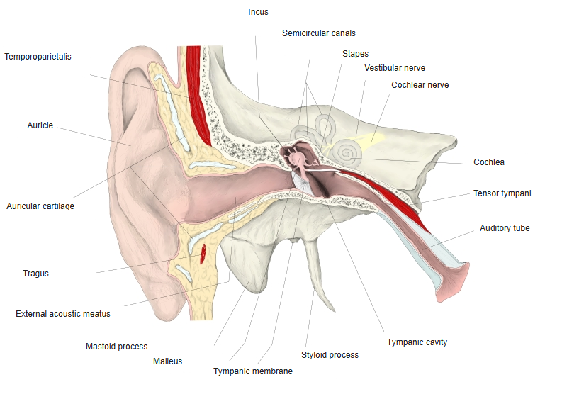
| External ear | Auricle, external acoustic meatus, tympanic membrane | Collects sound and guides it to the tympanic membrane |
| Middle ear | Tympanic cavity with the auditory ossicles, auditory tube, mastoid antrum and cells | Transmits and amplifies the vibrations of the tympanic membrane to the internal ear |
| Internal ear | Bony labyrinth and membranous labyrinth: cochlea, vestibule, semicircular canals | Hearing (cochlea) and balance (utricle, saccule, semicircular ducts) |
- The middle ear is an air-filled space; the internal ear is filled with fluid (perilymph and endolymph).
- The auditory tube connects the middle ear with the nasopharynx and equalises the air pressure on both sides of the tympanic membrane.
Right ear in the temporal bone (superior view) 
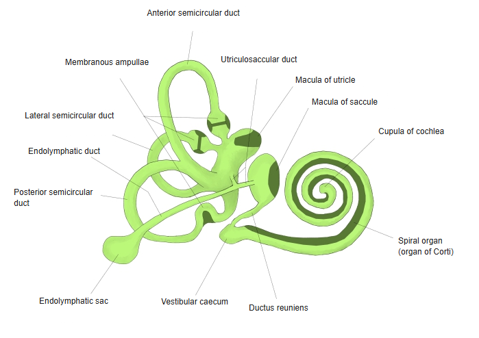
Membranous labyrinth
Pathway of sound
- The auricle collects sound waves and the external acoustic meatus carries them to the tympanic membrane, which vibrates.
- The chain of ossicles – malleus, incus and stapes – passes the vibration to the oval window.
- Because the tympanic membrane is much larger than the footplate of the stapes, the pressure is increased many times; this matches the sound in air to the fluid of the internal ear.
- Movements of the stapes set up waves in the perilymph of the cochlea; the basilar membrane vibrates and bends the hair cells of the spiral organ.
- The pressure wave is released through the round window back into the middle ear.
- The tensor tympani and stapedius reduce the movement of the ossicles in response to loud sound.
Balance
- The vestibular apparatus consists of the utricle, saccule and three semicircular ducts.
- The maculae of the utricle and saccule sense the position of the head and straight-line acceleration (gravity, lifts, cars).
- The ampullary crests of the semicircular ducts sense rotation of the head in any plane.
- Their signals travel in the vestibular nerve; those of the cochlea in the cochlear nerve.
References and sources
- Further reading: Ear (vestibulocochlear organ), Wikipedia.