Foramen spinosum (Latin: foramen spinosum)
The foramen spinosum is:
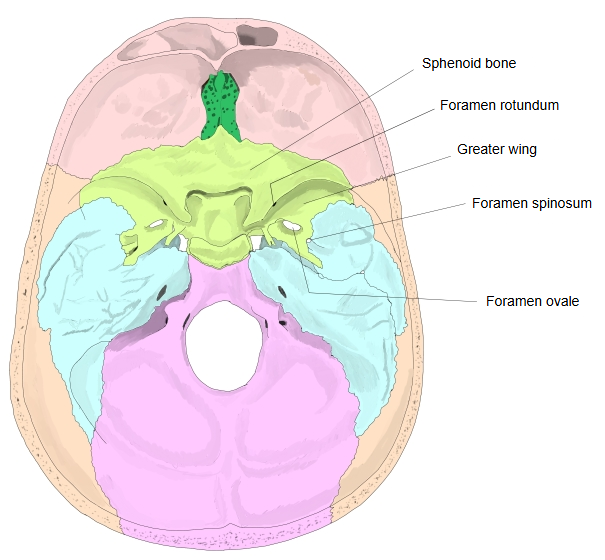
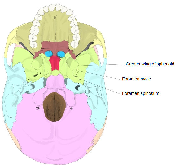
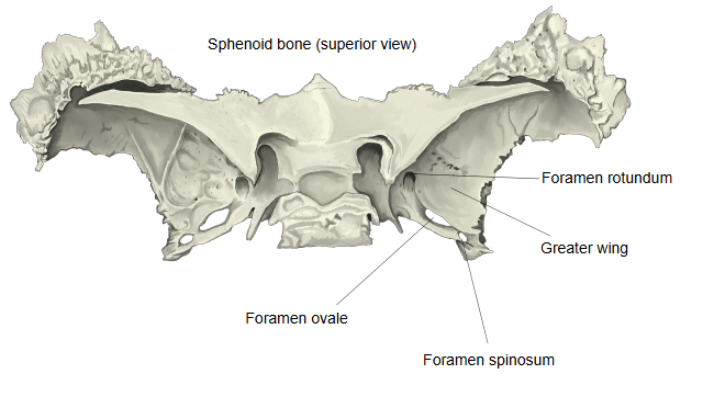
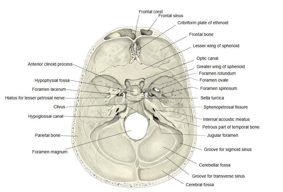
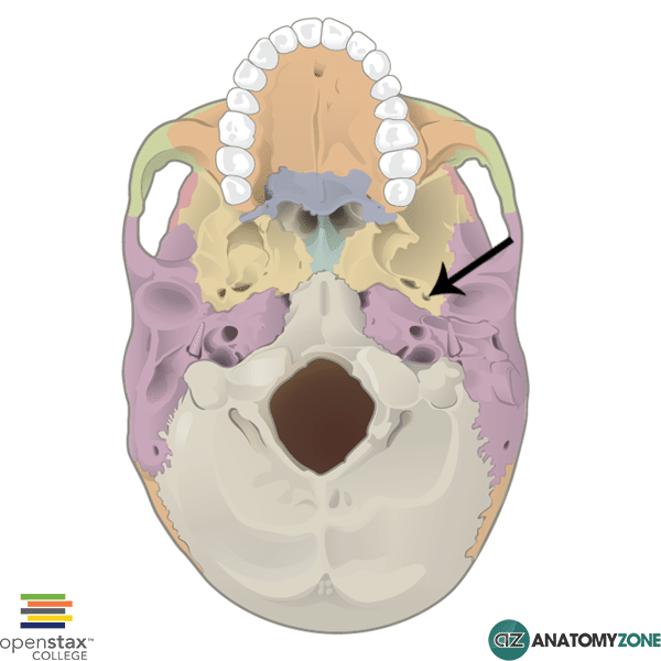
It lies in the floor of the middle cranial fossa, in front of the spine of the sphenoid, from which it takes its name. On the outer surface of the skull base it opens into the infratemporal fossa; the mandibular fossa of the temporal bone lies lateral to it, and the groove for the cartilaginous part of the auditory tube runs posteromedial to it. Inside the skull, the groove for the middle meningeal artery starts at this opening and branches over the inner surface of the temporal and parietal bones. The foramen rotundum and the foramen ovale are the other openings of the greater wing, further forward.
Nerve, artery and vein passing through it
The following vessels and nerve pass through the foramen spinosum.
| 1 | Nerve | · Meningeal branch of the mandibular nerve (the mandibular nerve is the third division of the trigeminal nerve); also called the nervus spinosus, it turns back into the skull to supply the dura mater. |
| 2 | Artery | · Middle meningeal artery (a branch of the maxillary artery); it supplies the dura mater and the bones of the skull vault. |
| 3 | Vein | · Middle meningeal veins, which run with the artery. |
Size and variation
· It becomes a complete bony ring in infancy or early childhood (reported from about 8 months to 7 years after birth).
· It may be incomplete, open toward the edge of the bone, in a large share of skulls; some studies give almost half.
· Rarely it is missing, usually on one side, and the middle meningeal artery then enters through the foramen ovale. Fewer than 1% of skulls have a double foramen, often with a doubled artery.
Note: the foramen spinosum is a landmark in skull-base surgery. Tearing of the middle meningeal artery, which runs from here over the inside of the skull, is the usual cause of an extradural (epidural) haematoma after a blow to the side of the head.
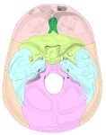 Internal cranial base
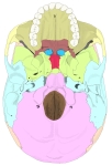 External cranial base
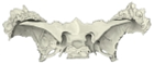 Sphenoid bone (superior view)
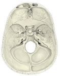 Internal cranial base
Reference websites
[Wikipedia]
· Foramen spinosum, Wikipedia.
· Middle meningeal artery, Wikipedia.
[Websites with illustrations]
· Foramen spinosum: anatomy and function (Kenhub)
· Foramen spinosum on the external skull base (AnatomyZone)
· Sphenoid bone, upper surface (Gray's Anatomy) (Wikimedia Commons)

· Human skull from below, with the foramen spinosum labelled (Alamy)

Where it is found
Related terms
- Foramen ovale
- Foramen rotundum
- Greater wing
- Infratemporal fossa
- Sphenoid bone
- Middle meningeal artery
- Middle meningeal veins
- Meningeal branch of mandibular nerve
References and sources
- Further reading: Foramen spinosum, Wikipedia.
- Further reading: Middle meningeal artery, Wikipedia.
- Federative International Programme for Anatomical Terminology (FIPAT). Terminologia Anatomica, 2nd ed. (TA2), 2019.