Tragus (Latin: tragus)
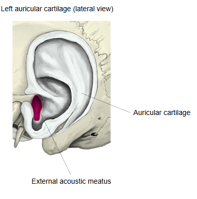
· It is a flat projection of cartilage in front of the external acoustic opening, and projects backwards over the opening of the external acoustic meatus. Above, the anterior notch separates it from the crus of the helix. It contains a plate of elastic cartilage (the tragal lamina), part of the auricular cartilage and continuous with the cartilage of the ear canal, and gives attachment to a small intrinsic muscle of the auricle, the tragicus (see auricular muscles). Its inner surface often carries hairs (tragi), which become more prominent with age; its name comes from the Greek word for goat, for this tuft of hair. Pressing the tragus closes the ear canal; pain on pressure suggests inflammation of the external ear canal (otitis externa).
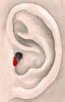 Left auricle (lateral surface)
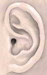 Parts of the auricle
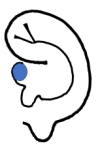 Auricle (diagram)
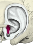 Left auricular cartilage (lateral surface)
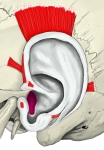 Intrinsic and extrinsic auricular muscles
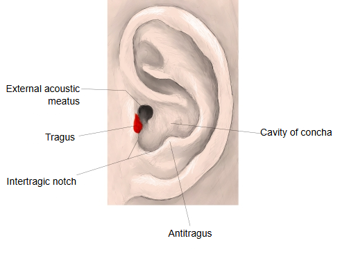
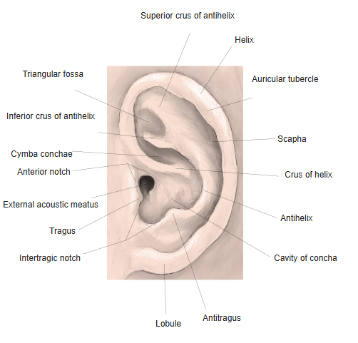
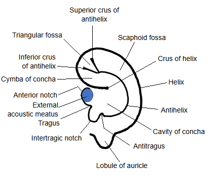
Names of the parts of the auricle
The surface of the auricle is full of ridges and hollows, and its parts have the following names.
Note: the antitragohelicine fissure can also be seen from the lateral surface.
Reference websites
[Wikipedia]
· Tragus (ear), Wikipedia.
· Auricle (anatomy), Wikipedia.
[Websites with illustrations]
· Outer ear (auricle) with its parts labelled (iStock)
· Ear anatomy infographic (iStock)
· The auricle, lateral surface (Gray's Anatomy, fig. 904) (Wikimedia Commons)

Related terms
- Antitragus
- Antitragohelicine fissure
- Helix
- Crus of helix
- Spine of helix
- Cauda helicis
- Intertragic notch
- Lobule of auricle
- Auricle
- External ear
- Concha of auricle
- Anterior notch of auricle
- Auricular cartilage
- Auricular muscles
- External acoustic meatus
References and sources
- Further reading: Tragus (ear), Wikipedia.
- Further reading: Auricle (anatomy), Wikipedia.
- Federative International Programme for Anatomical Terminology (FIPAT). Terminologia Anatomica, 2nd ed. (TA2), 2019.