Distal tubule (Latin: tubulus distalis)
In the narrow sense, the distal convoluted tubule
- Overview
- Straight and convoluted parts compared
- Reference websites
- (Wikipedia, websites with illustrations)
- Related terms
Overview
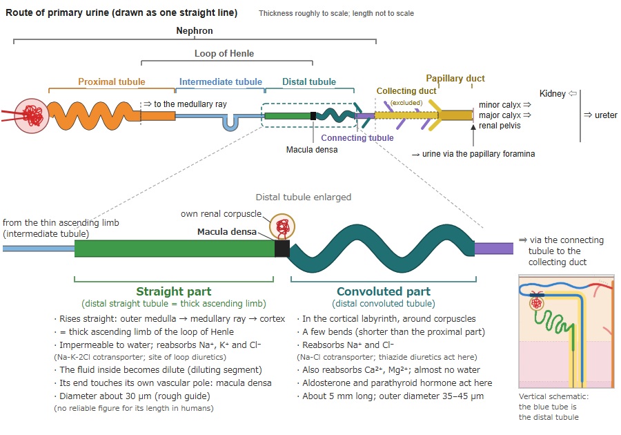
· It begins at the thin ascending limb of the intermediate tubule (in cortical nephrons with a short loop, at the thin descending limb).
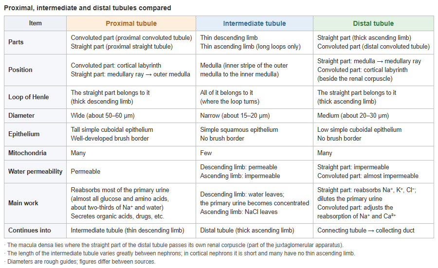
- Proximal tubule : follows Bowman's capsule (convoluted and straight parts); reabsorbs most of the water and nutrients in the primary urine. About 14 mm long.
- Intermediate tubule : the thin part of the loop of Henle (thin descending and thin ascending limbs); takes part in concentrating the urine in the medulla. Length varies.
- Distal tubule : straight part (thick ascending limb) and convoluted part; drains through the connecting tubule into the collecting duct and adjusts electrolytes and water under hormonal control.
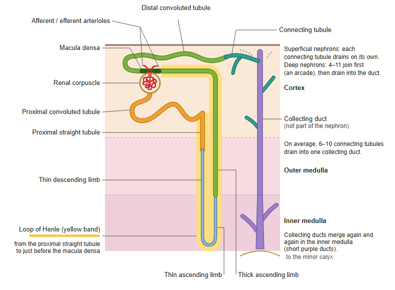
· In the nephron (renal corpuscle + renal tubule) it is the third division, after the proximal tubule and the intermediate tubule. Some sources use the name distal tubule only for the convoluted part (the distal convoluted tubule).
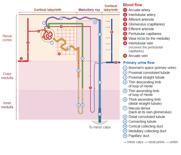
· It is divided into a straight part that runs straight up (the distal straight tubule = thick ascending limb, one of the straight tubules) and a convoluted part that winds (the distal convoluted tubule). The straight part rises from the outer zone of the renal medulla through the medullary rays to the cortex. The convoluted part lies in the cortical labyrinth of the renal cortex, where its sections are seen beside renal corpuscles and sections of the proximal tubule. The straight part is the last, ascending part of the loop of Henle.
· The end of the straight part, where it turns into the convoluted part, touches the vascular pole of its own renal corpuscle (the tuft of glomerular capillaries inside Bowman's capsule); here the macula densa forms. The macula densa is part of the juxtaglomerular apparatus.
· The epithelium is simple cuboidal. Its cells are lower than in the proximal tubule and have no brush border, so the lumen looks wide. The cells are rich in mitochondria and have deep infoldings at their base.
Straight and convoluted parts compared
· The straight part (thick ascending limb) does not let water through but reabsorbs sodium, potassium and chloride (Na-K-2Cl cotransporter). The fluid inside therefore becomes dilute, while the surrounding renal medulla becomes more concentrated. Loop diuretics act by blocking this cotransporter.
· The convoluted part (distal convoluted tubule) reabsorbs sodium and chloride (thiazide-sensitive Na-Cl cotransporter), secretes potassium and reabsorbs calcium (promoted by parathyroid hormone), and so helps regulate potassium, sodium, calcium and pH. Sodium reabsorption is increased by aldosterone. Thiazide diuretics act by blocking the Na-Cl cotransporter here.
· The table below summarizes the comparison.
| Item | Straight part (distal straight tubule = thick ascending limb) | Convoluted part (distal convoluted tubule) |
|---|---|---|
| Position | From the outer medulla through the medullary ray to the cortex | Cortical labyrinth of the renal cortex (around the renal corpuscle) |
| Shape | Rises almost straight | Starts just beyond the macula densa and bends a few times (shorter than the proximal convoluted part) |
| Length / diameter | Diameter about 30 µm (rough guide); no reliable figure for its length | About 5 mm long (rough guide); outer diameter about 35–45 µm |
| Epithelium | Simple cuboidal (low cells); no brush border | Simple cuboidal (lower than in the proximal tubule); no brush border, so the lumen looks wide |
| Cell base | Both have deep basal infoldings and many mitochondria | |
| Main work | Reabsorbs Na⁺, K⁺ and Cl⁻ (Na-K-2Cl cotransporter; about 25% of the filtered Na⁺); Ca²⁺ and Mg²⁺ are also reabsorbed between the cells | Reabsorbs Na⁺ and Cl⁻ (Na-Cl cotransporter); reabsorbs Ca²⁺ (increased by parathyroid hormone); reabsorbs Mg²⁺ |
| Water | Almost impermeable (the fluid becomes dilute: diluting segment) | Almost impermeable |
| Diuretics acting here | Loop diuretics (furosemide and others) | Thiazide diuretics |
| Special features | At its end lies the macula densa (part of the juxtaglomerular apparatus); makes uromodulin | In its later part, aldosterone increases Na⁺ reabsorption |
| Continues into | The convoluted part | Connecting tubule (→ collecting duct) |
| Loop of Henle | Included (thick ascending limb of the loop of Henle) | Not included |
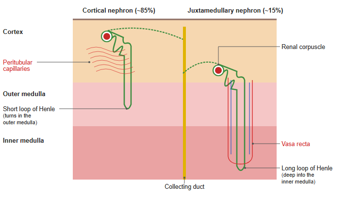
· In nephrons with a short loop (cortical nephrons) there is no thin ascending limb, and the straight part follows the thin descending limb. Some sources call only the convoluted part the distal tubule. Figures are rough guides and differ between sources.
→ Proximal, intermediate and distal tubules compared (table)
 Renal lobe and renal lobule
 Tissue organization of the renal cortex
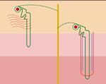 Cortical and juxtamedullary nephrons
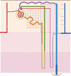 Nephron and blood vessels
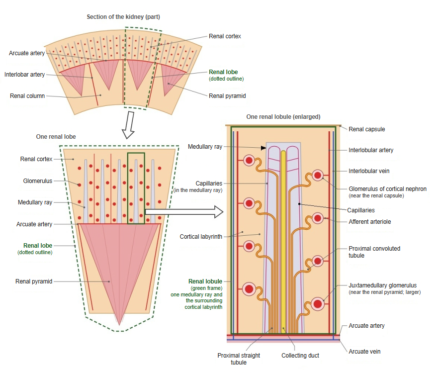
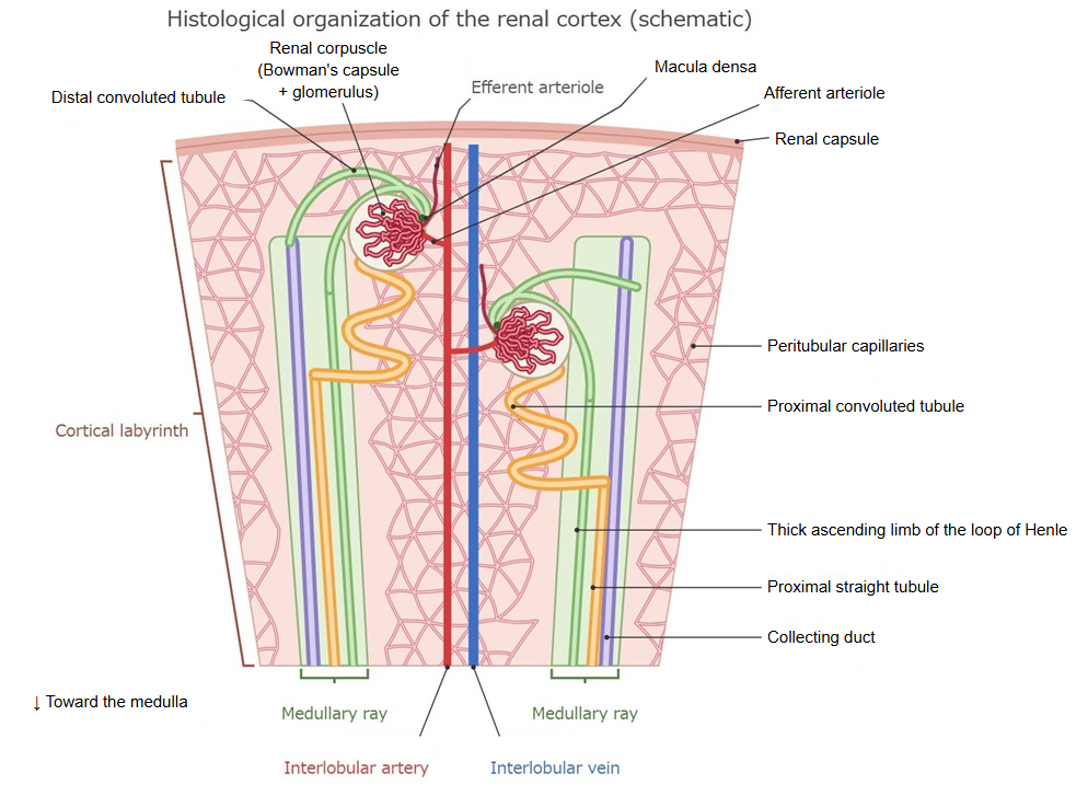
Reference websites
[Wikipedia]
· Distal convoluted tubule, Wikipedia.
· Nephron, Wikipedia.
[Websites with illustrations]
· Distal tubule (histology) (McGill University histology atlas)
· Kidney tubules (histology) (McGill University histology atlas)
· Tubules histology (kidneypathology.com)
· Kidney (virtual slide) (Histology Guide)
Related terms
- Juxtaglomerular apparatus
- Juxtaglomerular cells
- Macula densa
- Vascular pole
- Proximal tubule
- Straight tubule
- Cortical collecting duct
- Medullary collecting duct
- Papillary duct
- Kidney
- Renal parenchyma
- Renal pelvis
- Ureter
- Urinary system
References and sources
- Further reading: Distal convoluted tubule, Wikipedia.
- Further reading: Nephron, Wikipedia.
- Federative International Programme for Anatomical Terminology (FIPAT). Terminologia Anatomica, 2nd ed. (TA2), 2019.