Iliocostalis (Latin: musculus iliocostalis)
Also called the iliocostalis muscle
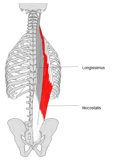
· It runs from the iliac crest and sacrum up the back of the ribs, attaching to the angles of the ribs, to the transverse processes of the lower cervical vertebrae. It lies immediately lateral to the longissimus.
· Acting on both sides it extends the vertebral column; acting on one side it bends the trunk to that side, and the lumbar part can also depress the lower ribs.
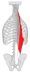 Iliocostalis and longissimus
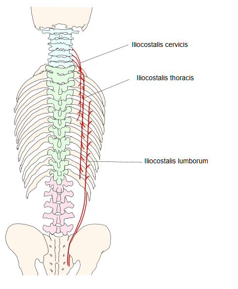
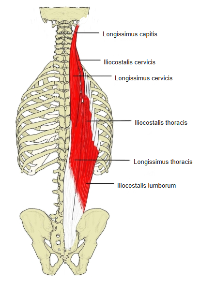
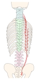 Three parts of the iliocostalis
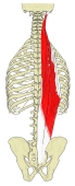 Erector spinae (lateral columns)
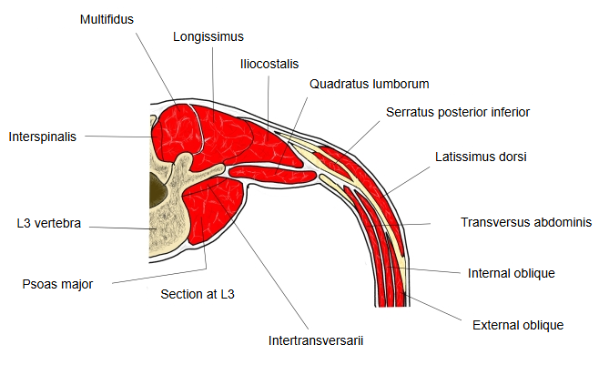
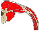 Back muscles at L3 (section)
The diagram below shows the classification of the muscles of the back.
- Superficial back muscles (5) : = spinohumeral muscles
- 1st layer (2)
- 2nd layer (3)
- Deep back muscles (16)
- 1st layer (2) : = spinocostal muscles
- 2nd layer (14)
- Muscles of the vertebral column (10) : = intrinsic back muscles
- Splenius muscles (2)
- Erector spinae (3) : iliocostalis, longissimus, spinalis
- Transversospinalis muscles (3)
- Others (2)
- Suboccipital muscles (4)
- Muscles of the vertebral column (10) : = intrinsic back muscles
Among the intrinsic muscles, the muscles of the vertebral column include the splenius muscles, the erector spinae (iliocostalis, longissimus and spinalis) and the transversospinalis group. In a cross-section at L3, the iliocostalis lies lateral to the longissimus, enclosed with it by the thoracolumbar fascia and behind the quadratus lumborum.
- Iliocostalis cervicis : from the angles of the 3rd–6th ribs to the posterior tubercles of the transverse processes of C4–C6.
- Iliocostalis thoracis : from the angles of the lower six ribs to the angles of the upper six ribs and the transverse process of C7.
- Iliocostalis lumborum : from the common erector spinae tendon (sacrum and iliac crest) to the angles of the lower six to eight ribs.
Origin: see the pages of the iliocostalis cervicis, iliocostalis thoracis and iliocostalis lumborum.
Insertion: see the pages of the iliocostalis cervicis, iliocostalis thoracis and iliocostalis lumborum.
Nerve: the lateral branches of the dorsal rami of the spinal nerves (about C8–L1).
Blood supply: the dorsal branches of the posterior intercostal arteries and the lumbar arteries.
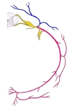 Thoracic nerve (scheme)
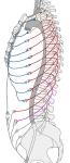 Intercostal arteries
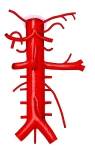 Abdominal aorta
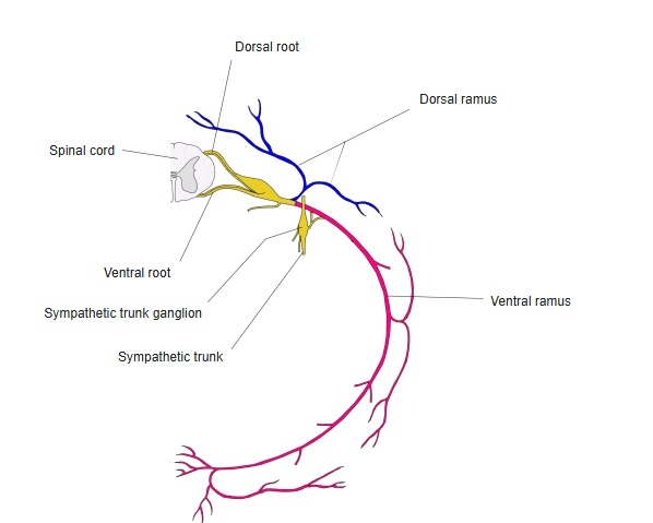
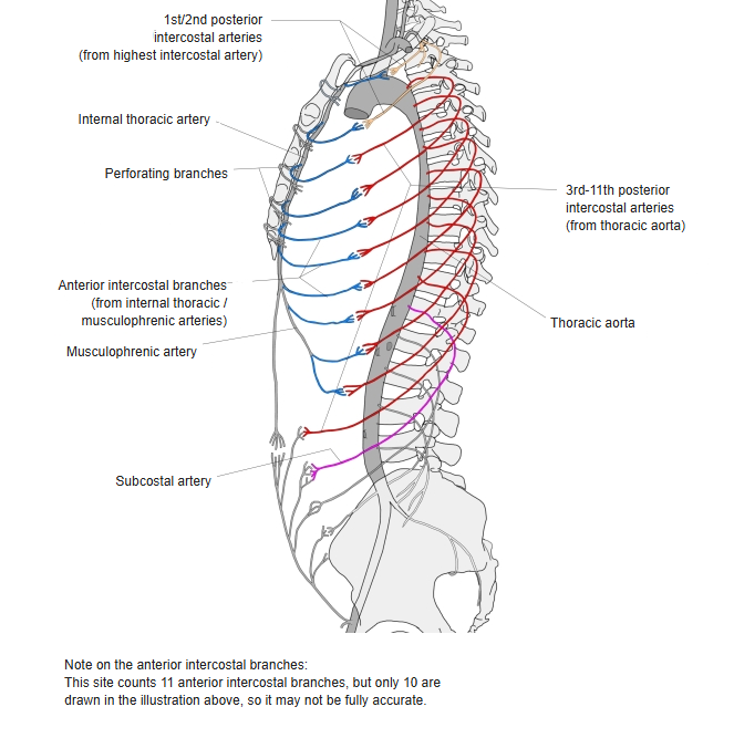
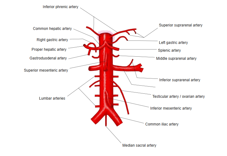
The cervical part separates fairly easily, but the lumbar and thoracic parts are hard to divide. Small extra muscle slips running from rib to rib are often found under the middle of the iliocostalis, especially in the 6th–8th intercostal spaces. They are thought to be remnants of the segmental pattern of the back muscles. They are supplied by the dorsal ramus (lateral branch) of the spinal nerve one segment higher, which suggests that they moved downwards during evolution.
Reference websites
[Wikipedia]
· Iliocostalis, Wikipedia.
· Erector spinae muscles, Wikipedia.
[Websites with illustrations]
· The intrinsic back muscles (TeachMeAnatomy)
· Deep muscles of the back (Gray's Anatomy, fig. 389) (Wikimedia Commons)

Related terms
- Erector spinae
- Iliocostalis lumborum
- Iliocostalis thoracis
- Iliocostalis cervicis
- Intrinsic back muscles
- Spinalis
- Thoracolumbar fascia
- Angle of rib
References and sources
- Further reading: Iliocostalis, Wikipedia.
- Further reading: Erector spinae muscles, Wikipedia.
- Federative International Programme for Anatomical Terminology (FIPAT). Terminologia Anatomica, 2nd ed. (TA2), 2019.