Latin: vertebrae cervicales · C1–C7
Overview
- The vertebral column is made up of five kinds of vertebrae; the seven cervical vertebrae form its uppermost part, in the neck.
- C1 – atlas: very different in shape from the other cervical vertebrae (no body and no spinous process).
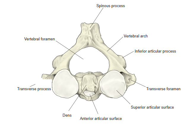
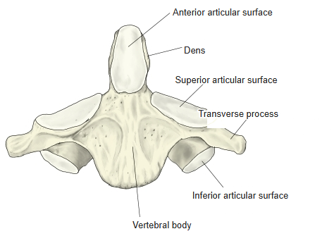
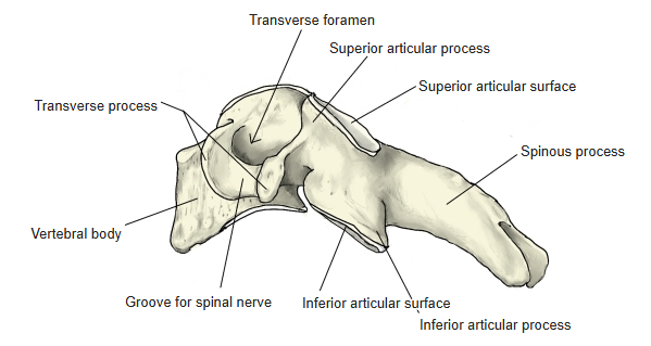
- C2 – axis: differs a little from a typical cervical vertebra; its main feature is the dens.
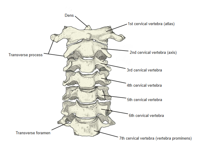
- C3–C7: typical cervical vertebrae; C7 is the vertebra prominens.
- The transverse foramina transmit the vertebral artery and vein; the artery usually enters at C6, so the foramen of C7 usually transmits only the vein.
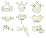
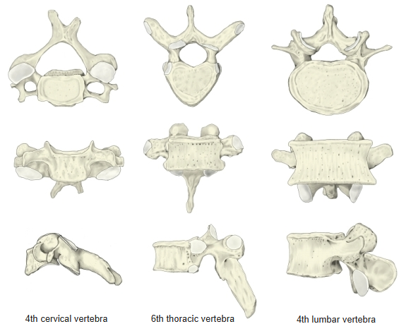
Cervical, thoracic and lumbar vertebrae compared
- Bones of the trunk
- Vertebral column
- Cervical vertebrae (7)
- Thoracic vertebrae (12)
- Lumbar vertebrae (5)
- Sacrum (1)
- Coccyx (1)
- Thoracic cage
- Sternum (1)
- Ribs (24)
- Vertebral column
Illustrations
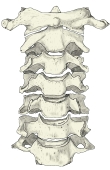
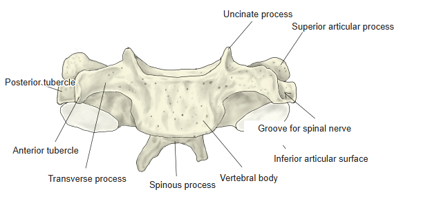
Cervical vertebrae (anterior) 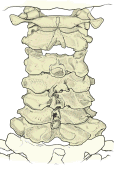
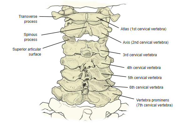
Cervical vertebrae (posterior) 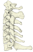
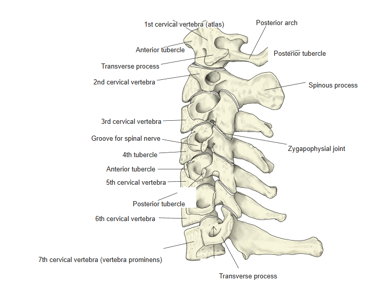
Cervical vertebrae (left lateral) 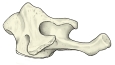
Atlas (left lateral) 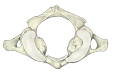
Atlas (from above) 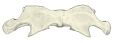
Atlas (anterior) 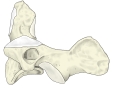
Axis (left lateral) 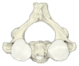
Axis (from above) 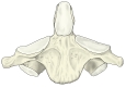
Axis (anterior) 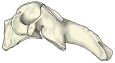
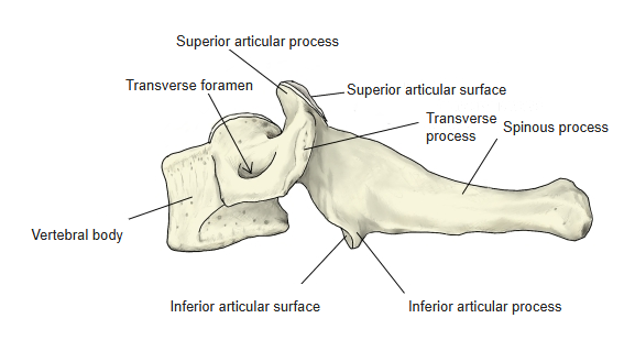
4th cervical vertebra (left lateral) 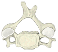
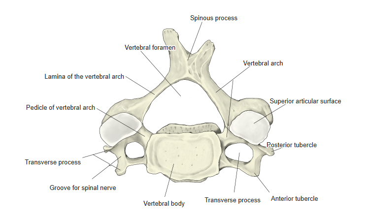
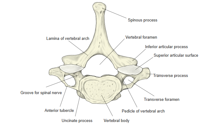
4th cervical vertebra (from above) 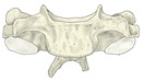
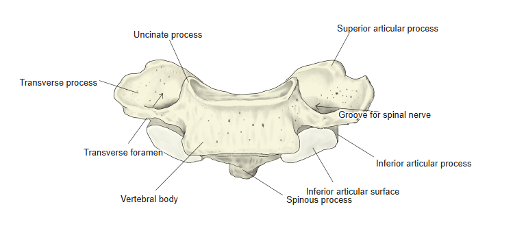
4th cervical vertebra (anterior) 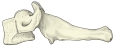
7th cervical vertebra (left lateral) 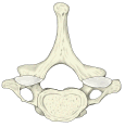
7th cervical vertebra (from above) 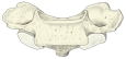
7th cervical vertebra (anterior)
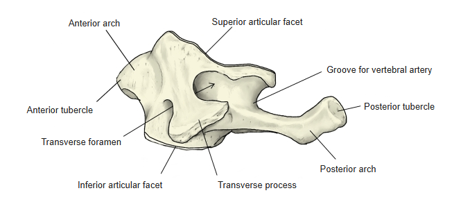
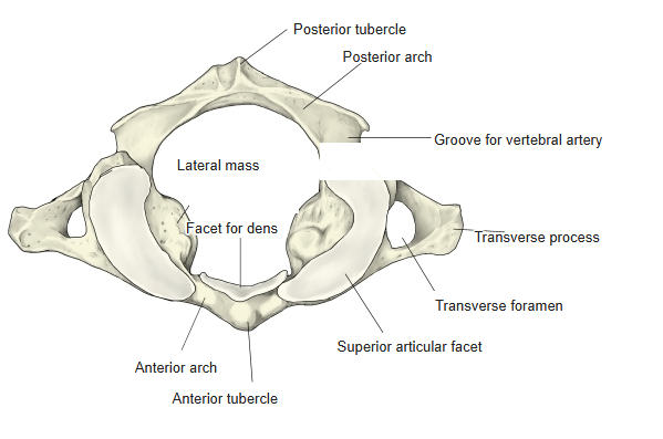
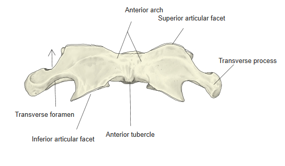
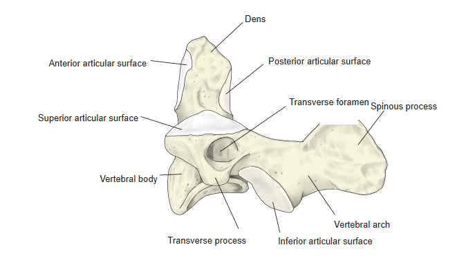
Articulations
| # | Bone | Joint |
|---|---|---|
| 1 | Atlas (superior articular facets) ⇔ occipital bone (occipital condyles) | Atlanto-occipital joint |
| 2 | Atlas (facet for dens) ⇔ axis (dens, anterior articular surface) | Median atlanto-axial joint |
| 3 | Atlas (inferior articular facets) ⇔ axis (superior articular surfaces) | Lateral atlanto-axial joint |
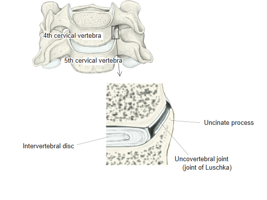
| 4 | Uncinate process ⇔ lateral edge of the body of the vertebra above | Uncovertebral joints |
| 5 | Inferior articular surface ⇔ superior articular surface of the next vertebra | Zygapophysial (facet) joints |
| 6 | Vertebral bodies (C7 with C6 and T1, etc.) | Intervertebral joints (symphyses) |
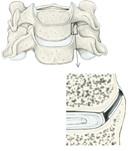
Uncovertebral joint
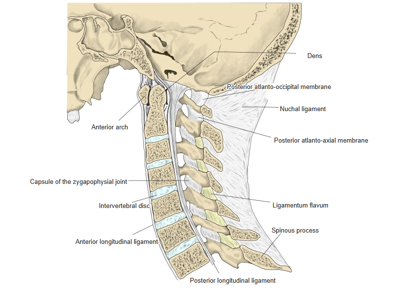
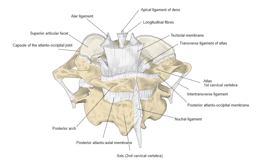
Ligaments
The following ligaments attach to the cervical vertebrae. The list is intended to be comprehensive but may omit minor ligaments.
| # | Ligament | Connects |
|---|---|---|
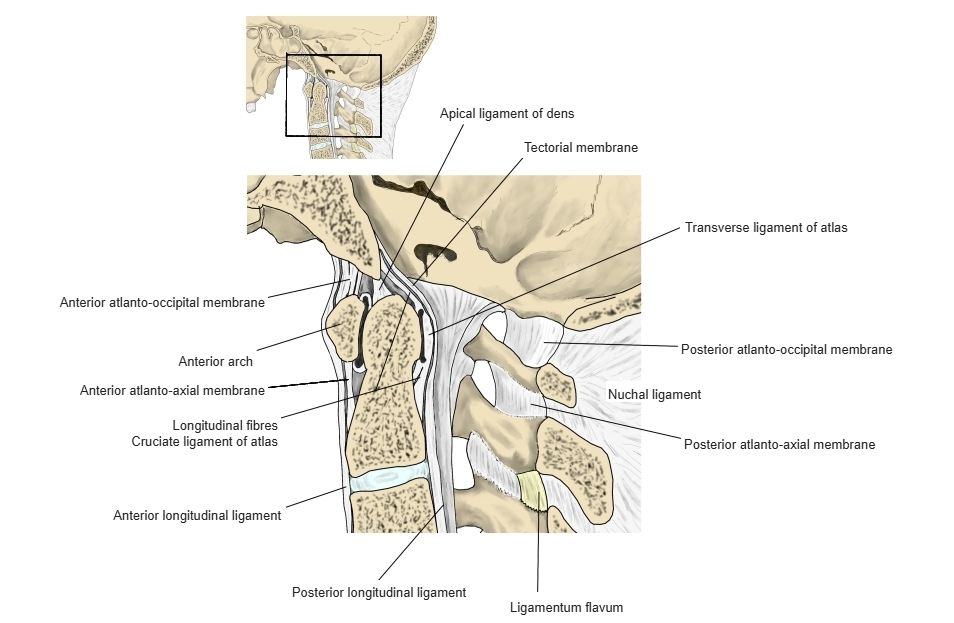
| 1 | Apical ligament of dens | Apex of the dens – front of the foramen magnum (occipital bone) |
| 2 | Alar ligaments | Sides of the apex of the dens – medial sides of the occipital condyles |
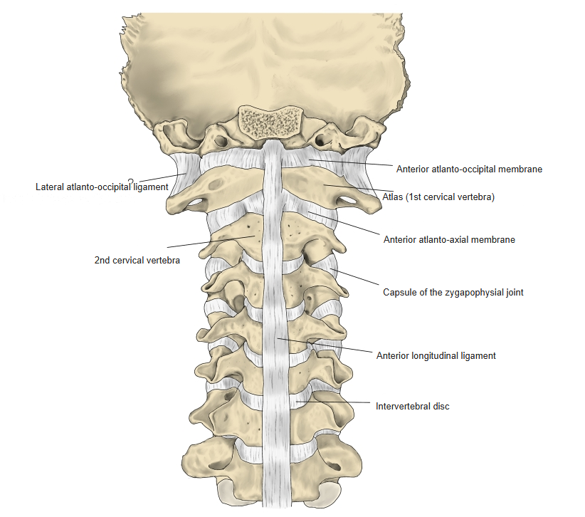
| 3 | Anterior longitudinal ligament | Occipital bone (pharyngeal tubercle) – front of the sacrum; attaches to the anterior tubercle of the atlas |
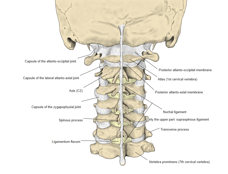
| 4 | Nuchal ligament | Occipital bone – spinous processes of the cervical vertebrae |
| 5 | Intertransverse ligaments | Between the transverse processes |
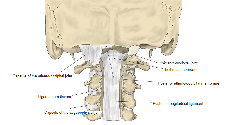
| 6 | Posterior longitudinal ligament | Occipital bone (clivus) – sacral canal |
| 7 | Ligamenta flava | Between the laminae |
| 8 | Longitudinal bands (of the cruciate ligament) | Back of the body of the axis – front of the foramen magnum |
| 9 | Posterior atlanto-axial membrane | Arch of the axis – posterior arch of the atlas |
| 10 | Transverse ligament of atlas (part of the cruciate ligament) | Between the lateral masses of the atlas |
| 11 | Lateral atlanto-occipital ligament | Occipital bone – transverse process of the atlas |
| 12 | Anterior atlanto-occipital membrane | Occipital bone – anterior arch of the atlas |
| 13 | Posterior atlanto-occipital membrane | Occipital bone – posterior arch of the atlas |
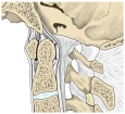
Ligaments of the craniovertebral junction (median section) 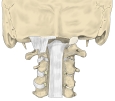
Ligaments (posterior, vertebral canal opened) 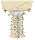
Ligaments of the cervical spine (posterior) 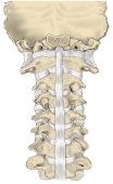
Ligaments of the cervical spine (anterior) 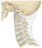
Ligaments of the cervical spine (median section) 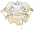
Ligaments of the atlas and axis (posterior)
Muscle attachments
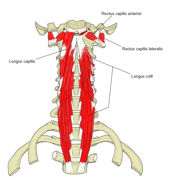
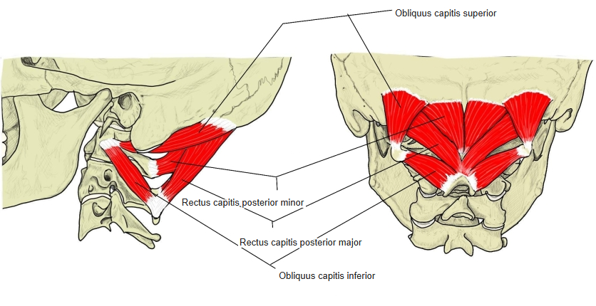
- Origin of: Rectus capitis posterior minor, Rectus capitis posterior major, Obliquus capitis inferior, Obliquus capitis superior, Rectus capitis lateralis, Rectus capitis anterior, Levator scapulae, Spinalis cervicis, Interspinales cervicis, Rhomboid minor, Serratus posterior superior, Splenius capitis, Trapezius, Intertransversarii, Longus capitis, Longus colli, Scalenus anterior, Scalenus medius, Scalenus posterior, Semispinalis capitis
- Insertion of: Iliocostalis, Splenius cervicis, Semispinalis cervicis
Muscles that both arise from and insert on cervical vertebrae are listed under origin. Splenius capitis, trapezius and serratus posterior superior arise from the nuchal ligament and lower cervical spinous processes.
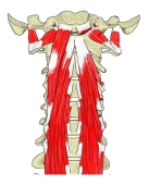
Prevertebral muscles (anterior) 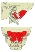
Suboccipital muscles (posterior)
Parts of a cervical vertebra
- Vertebral body – larger in the lower vertebrae, but smaller than in the thoracic and lumbar vertebrae.
- Transverse process – one of the most characteristic features: much broader and shorter than in other vertebrae, with a transverse foramen for the vertebral artery and vein.
- Anterior tubercle – the thickened end in front of the transverse foramen. The anterior tubercle of C6 can be palpated through the skin just behind the common carotid artery and is called the carotid tubercle; it is a landmark for compressing the artery and for locating the vertebral artery.
- Posterior tubercle – the thickened end behind the transverse foramen.
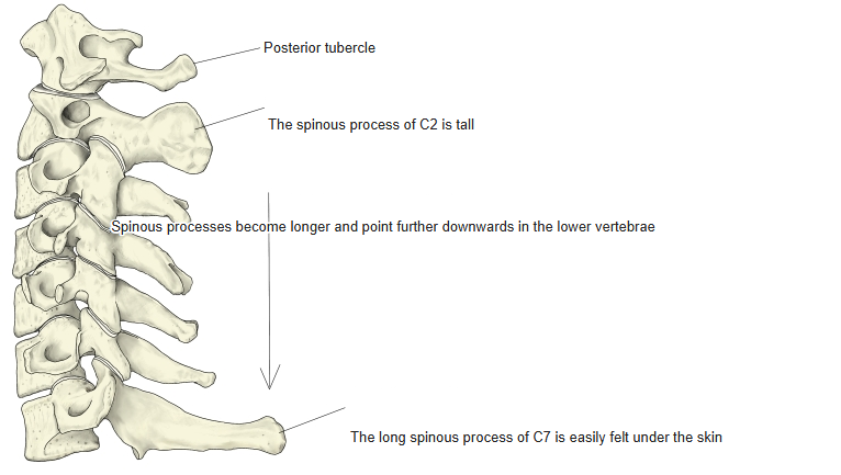
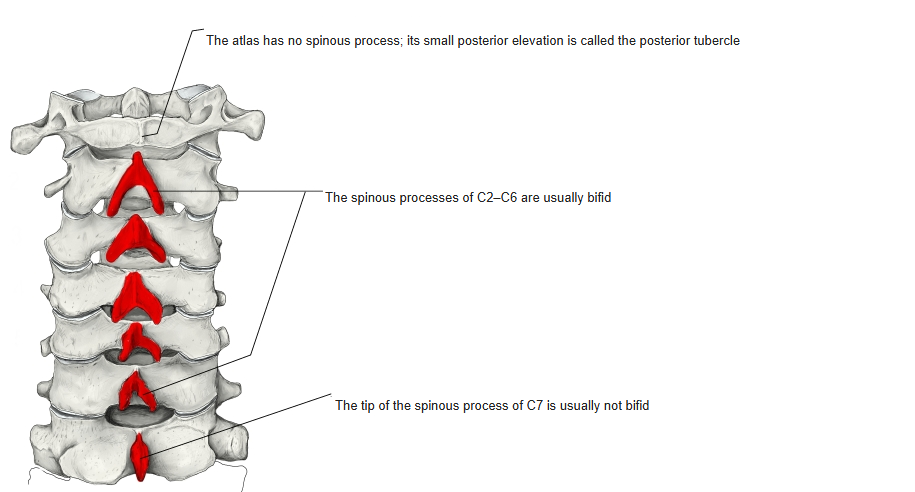
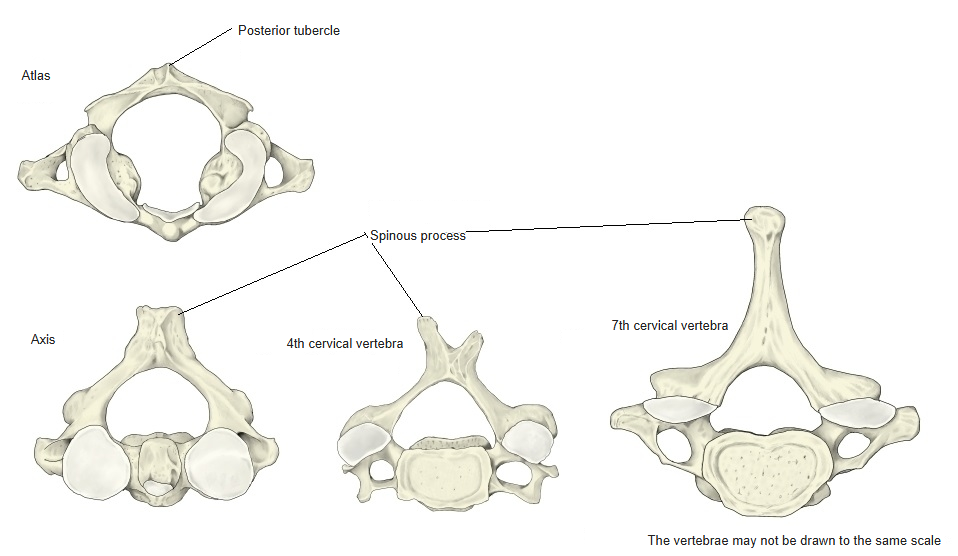
- Spinous process – absent in the atlas (its place is taken by a small posterior tubercle); tall in the axis; bifid from C2 to C6 (C6 less clearly); longer and pointing more downwards in the lower vertebrae; the long, usually undivided spinous process of C7 is easily felt under the skin.
- Vertebral foramen – large and triangular.
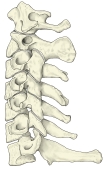
Spinous processes (lateral) 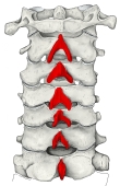
Bifid spinous processes (posterior) 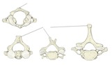
Spinous processes of C1, C2, C4 and C7
References and sources
- Further reading: Cervical vertebrae, Wikipedia.