Maxillary hiatus (Latin: hiatus maxillaris)
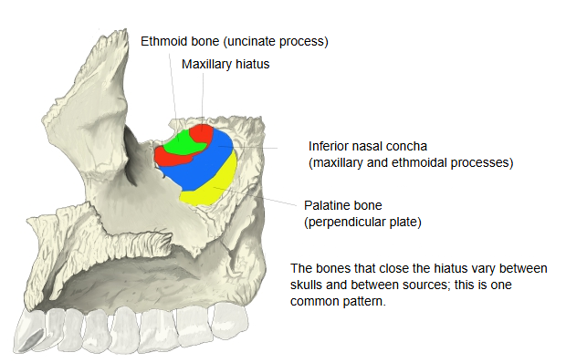
· In the complete skull most of it is covered by other bones: the palatine bone, the ethmoid bone and the inferior nasal concha. What remains, together with the mucosa, is the small opening of the maxillary sinus, which lies high on the medial wall of the sinus and opens into the semilunar hiatus of the middle nasal meatus.
The usual pattern is closure by the ethmoid and palatine bones (with the inferior nasal concha), but an image search shows several different patterns; the exact arrangement varies.
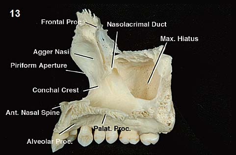
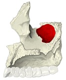 Maxilla (medial view)
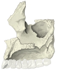 Maxilla (medial view, labelled)
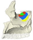 How the hiatus is closed
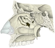 Lateral wall of nasal cavity
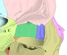 Right orbit, cut open (lateral view)
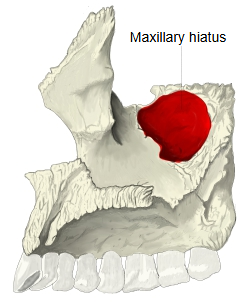
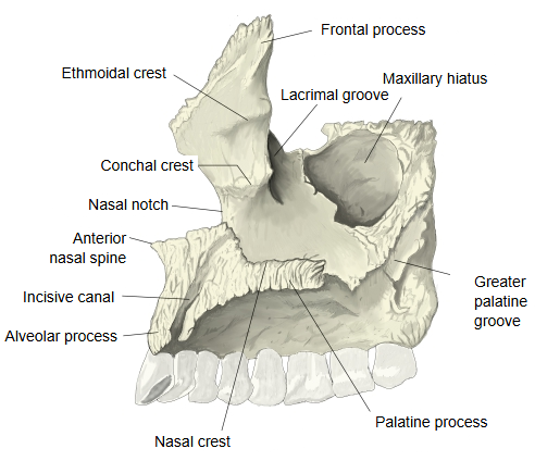
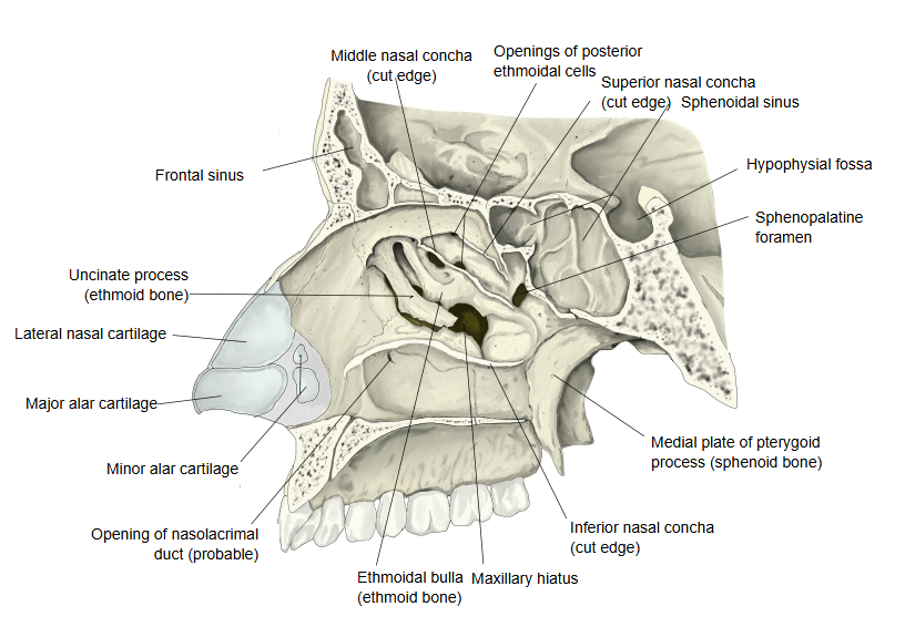
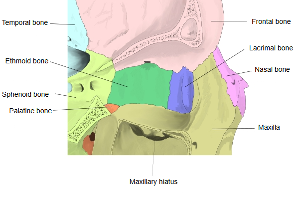
It is a large gap in the upper back part of the nasal surface, the exit of the maxillary sinus into the nasal cavity. In front of it the lacrimal groove runs downwards; with the lacrimal bone and the inferior nasal concha it forms the wall of the nasolacrimal canal. The back edge of the groove meets the lacrimal bone as the lacrimal margin. Small depressions above and behind the hiatus meet the ethmoidal labyrinth and the orbital process of the palatine bone; behind it, a rough area meets the perpendicular plate of the palatine bone.
On the isolated maxilla the hiatus is a large, roughly inverted-triangular opening in the upper back part of the nasal surface, the entrance to the maxillary sinus; in the complete skull it is largely closed from the medial (nasal) side by the palatine bone, the ethmoid bone and the inferior nasal concha. Its upper margin shows many small depressions, which complete the lateral walls of some of the ethmoidal air cells. Because the sinus opening lies high on its medial wall, the maxillary sinus does not drain by gravity when the head is upright.
Reference websites
[Wikipedia]
· Maxillary sinus, Wikipedia.
· Maxilla, Wikipedia.
[Websites with illustrations]
· Maxilla, medial view (photograph, maxillary hiatus labelled) (Neupsy Key)
· Maxillary sinus opened from the side (Gray's Anatomy, fig. 192) (Wikimedia Commons)

· Lateral wall of the nasal cavity with the maxillary hiatus (Jaypee Digital)

Where it is found
Related terms
- Maxillary sinus
- Body of maxilla
- Middle nasal meatus
- Uncinate process
- Nasolacrimal canal
- Perpendicular plate
References and sources
- Further reading: Maxillary sinus, Wikipedia.
- Further reading: Maxilla, Wikipedia.
- Federative International Programme for Anatomical Terminology (FIPAT). Terminologia Anatomica, 2nd ed. (TA2), 2019.