Fauces (Latin: fauces)
- Overview
- Muscles of the palate and fauces
- Vessels and nerves
- Palatine tonsil
- Reference websites
- (Wikipedia, websites with illustrations)
- Related terms
Overview
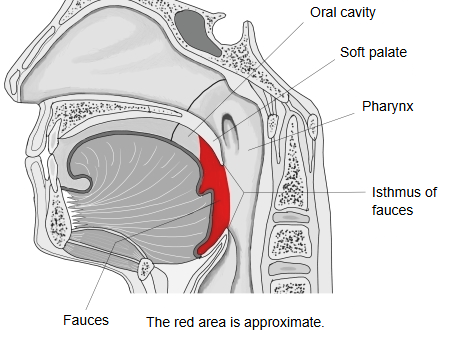
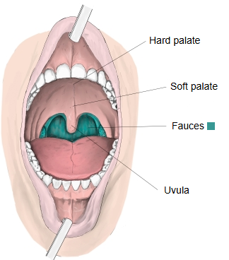
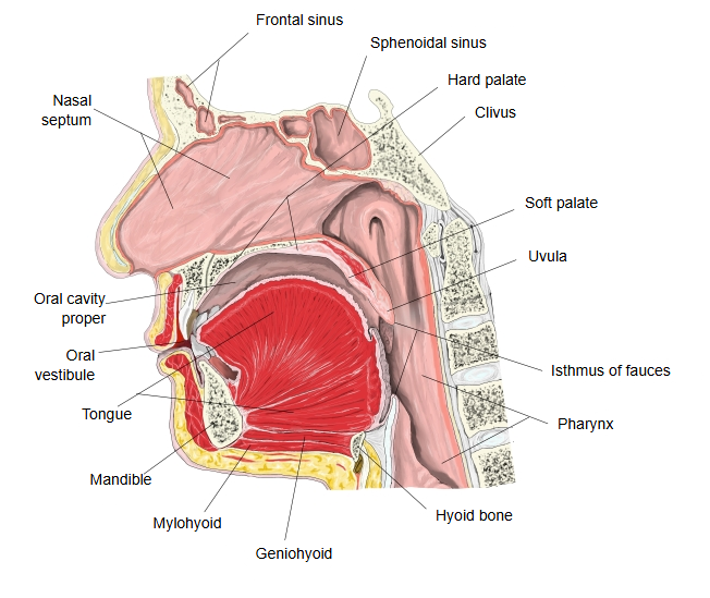
· It is the short passage at the back of the mouth that leads from the oral cavity into the pharynx.
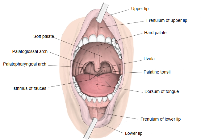
· Isthmus of fauces (the fauces in the narrow sense): the narrowest part of the fauces, at the palatoglossal arches; it marks the boundary between the mouth and the pharynx.
of the
fauces
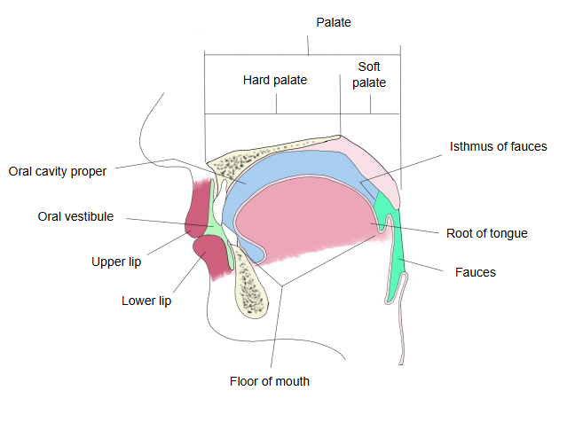
- Upper wall: soft palate : the back part of the palate, a plate of muscle covered with mucous membrane; the uvula hangs from its free edge.
- Lateral walls: palatine arches : two arch-shaped folds on each side, running down from the soft palate: the palatoglossal arch in front and the palatopharyngeal arch behind (the pillars of the fauces). The tonsillar fossa between them holds the palatine tonsil.
- Lower wall: root of tongue : the rearmost of the three parts of the tongue (apex, body and root).
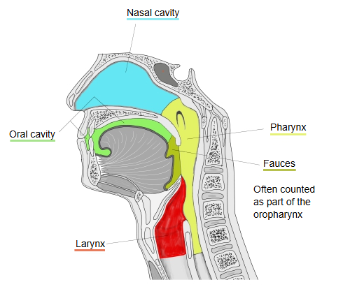
In that view the fauces is the subdivision of the oropharynx directly behind the oral cavity. Sources differ in how the name is used: some treat "fauces" and "isthmus of fauces" as the same opening, others count the fauces as the front part of the oropharynx.
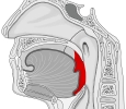 Fauces (median section)
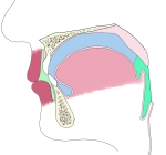 Oral cavity (diagram, median section)
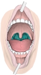 Fauces (anterior view)
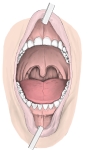 Oral cavity (anterior view)
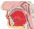 Oral cavity (median section)
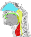 Nasal cavity, mouth, pharynx and larynx (median section)
Muscles of the palate and fauces
Five muscles belong to the palate and the fauces (the palatine muscles):
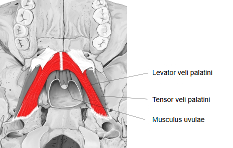
1. Levator veli palatini: a broad, rounded muscle. Action: raises the soft palate during swallowing, closing off the nasopharynx.
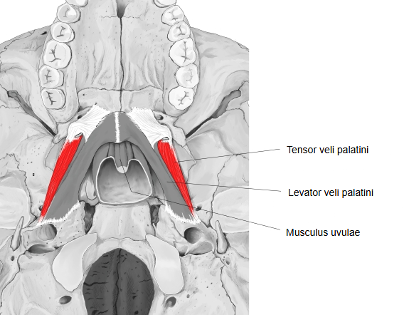
2. Tensor veli palatini: lies in front of and lateral to the levator veli palatini; its tendon turns around the pterygoid hamulus. Action: tightens the soft palate sideways; it also opens the auditory tube.
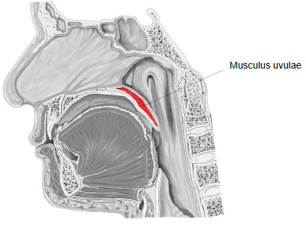
3. Musculus uvulae: lies inside the uvula. Action: shortens and lifts the uvula.
4. Palatoglossus: forms the palatoglossal arch. Action: raises the back of the tongue and draws the arches together, narrowing the fauces; this helps to control the movement of food in swallowing.
5. Palatopharyngeus: forms the palatopharyngeal arch. Action: pulls the soft palate down and raises the pharynx and larynx.
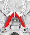 Levator veli palatini
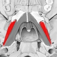 Tensor veli palatini
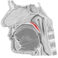 Musculus uvulae
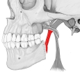 Palatoglossus
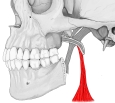 Palatopharyngeus
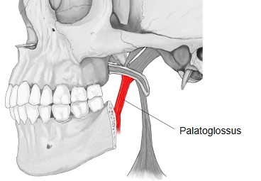
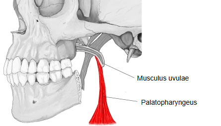
Vessels and nerves
Arteries
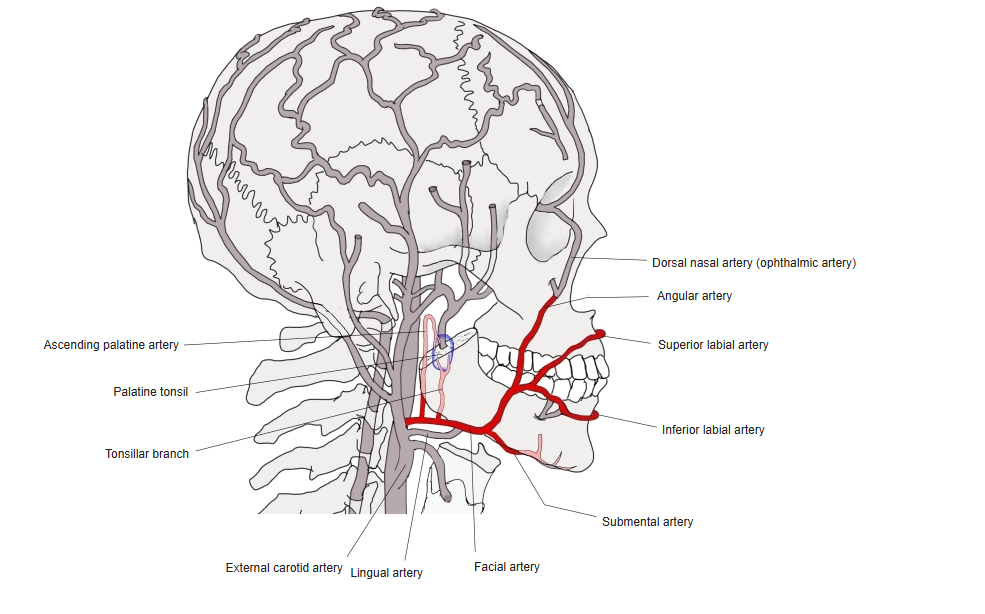
The soft palate is supplied by the descending palatine artery (through its lesser palatine branches), a branch of the pterygopalatine part of the maxillary artery, and by the ascending palatine artery, a branch of the facial artery.
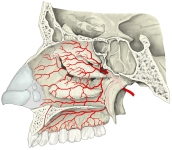 Branches of the maxillary artery
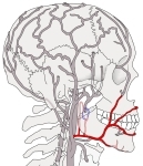 Facial artery
Veins
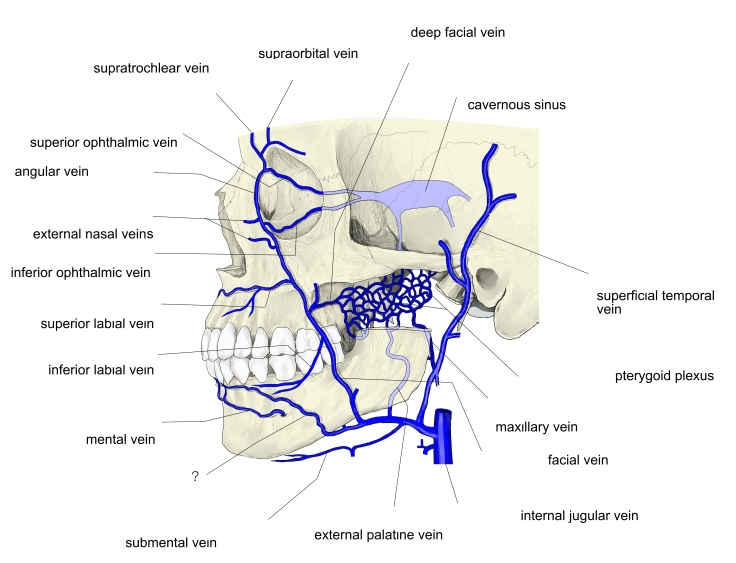
The veins of the soft palate drain mainly into the pterygoid plexus, which empties into the retromandibular vein.
Nerves
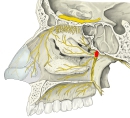
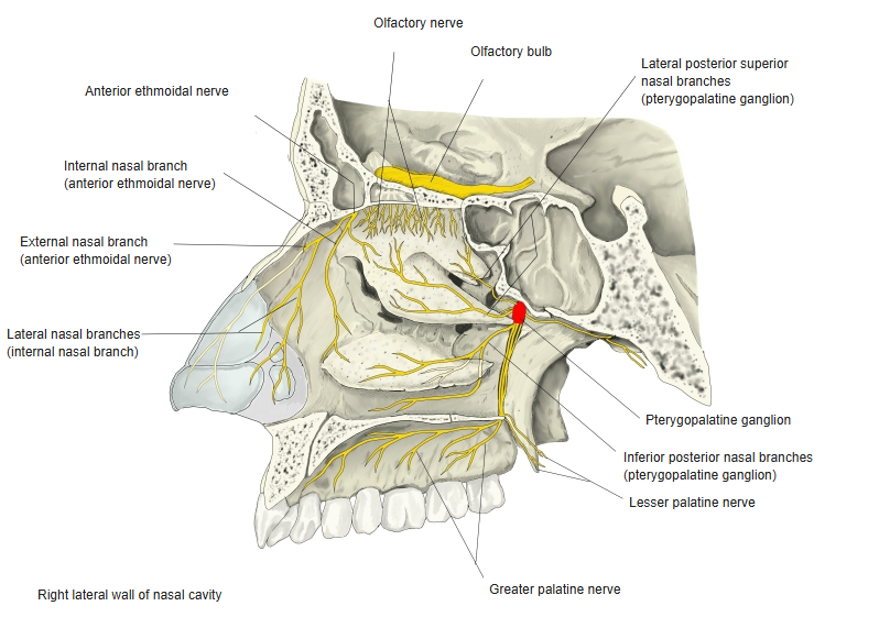
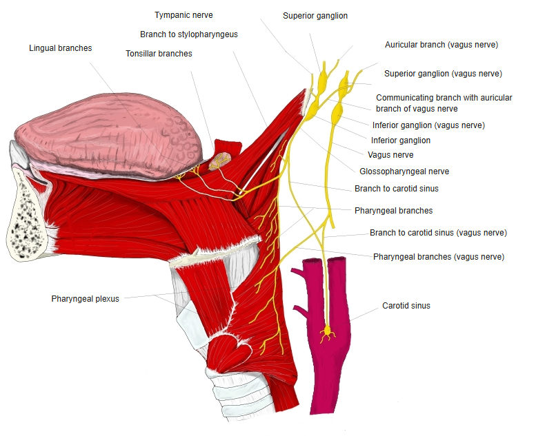
Sensory: the soft palate is supplied by the lesser palatine nerves, branches of the palatine nerves from the maxillary nerve (the second division of the trigeminal nerve); the glossopharyngeal nerve supplies the region of the palatine arches and tonsil.
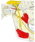
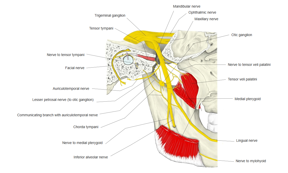
Motor: of the palatine muscles, the tensor veli palatini is supplied by the mandibular nerve (the third division of the trigeminal nerve); all the others, including the levator veli palatini, are supplied by the pharyngeal plexus
(formed by branches of the glossopharyngeal nerve and the vagus nerve; its motor fibres come mainly from the vagus).
 Pterygopalatine ganglion and palatine nerves
 Mandibular nerve
 Glossopharyngeal and vagus nerves
Palatine tonsil
⇒ See the entry palatine tonsil.
Reference websites
[Wikipedia]
· Fauces (throat), Wikipedia.
[Websites with illustrations]
· The palate (Kenhub)
· The oral cavity (TeachMeAnatomy)
Related terms
- Isthmus of fauces
- Soft palate
- Palatoglossal arch
- Palatopharyngeal arch
- Palatine tonsil
- Oropharynx
- Palatine muscles
References and sources
- Further reading: Fauces (throat), Wikipedia.
- Federative International Programme for Anatomical Terminology (FIPAT). Terminologia Anatomica, 2nd ed. (TA2), 2019.