Mucus
- Overview
- Main cells that secrete mucin, the main component of mucus
- Main sites where mucus is secreted
- Questions and answers
- Differences in the proportion and properties of mucin by site (examples)
- Key differences
- Note: classification of mucins
- Main contents of the secretory granules
- Where does the water in mucus come from?
- Mucin and water: mucin "draws in" water
- How the water comes out (schematic)
- Note: factors that regulate the water supply
- Related terms
Overview
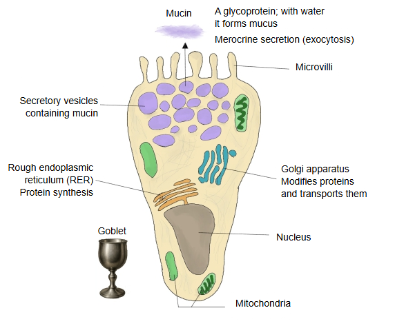
· Protection: shields the mucosa from acid, enzymes, microbes and mechanical irritation (friction).
· Lubrication: keeps the surface of the gastrointestinal tract and the respiratory tract smooth and helps the passage of food and air.
· Immune functions: traps particles and microbes, which are then removed by coughing, sneezing, defecation and so on.
· pH buffering: keeps the local environment stable (e.g. the mucus of the stomach).
Main cells that secrete mucin, the main component of mucus
A short list of the cells that secrete mucus:
| Cell | Main sites | Features and secretion | |
|---|---|---|---|
| 1 | Goblet cells | Epithelium of the gastrointestinal tract (especially the small and large intestine) and of the airways | Single cells that secrete mucin, stored in secretory vesicles and released by exocytosis (merocrine secretion). PAS-positive. Scattered in the epithelium. |
| 2 | Mucous gland cells | Salivary glands (part of the submandibular gland, the sublingual gland and others), glands of the airways (tracheal, bronchial and nasal glands) | Form multicellular glands. Pale, foamy cytoplasm; secrete mucin. The nucleus is flattened at the base of the cell. |
| 3 | Gland cells of the uterine cervix | Mucosa of the cervix of the uterus | Secrete cervical mucus, whose viscosity changes between the ovulatory and the luteal phase. |
| 4 | Cells of the duodenal glands (Brunner's glands) | Submucosa of the duodenum | Secrete alkaline mucus that protects the intestinal wall from gastric acid. |
| 5 | Mucous cells of the paranasal sinuses and the nasal cavity | Mucosa of the nasal cavity and the upper respiratory tract | The mucus traps particles and microbes in the inhaled air. Goblet cells and glands work together. |
| 6 | Surface mucous cells | Surface of the stomach | Cover the stomach lining with a protective layer of mucus. |
Main sites where mucus is secreted
A short list of the places where mucus is secreted:
| Site | Main role | Secretory cells | |
|---|---|---|---|
| 1 | Respiratory system (nasal cavity to trachea and bronchi) | Traps and removes particles and pathogens (swept out by the cilia) | Goblet cells, gland cells of the airways |
| 2 | Gastrointestinal tract (mouth to anus) | Lubrication; protects the mucosa from acid and digestive enzymes; eases mechanical irritation | Goblet cells, surface mucous cells, intestinal glands |
| 3 | Eye (conjunctiva, tear film) | Keeps the surface moist; removes particles | Goblet cells (conjunctiva), mucin-producing cells of the lacrimal glands |
| 4 | Ear (middle ear, auditory tube) | Protects the mucosa; prevents the entry of bacteria | Goblet cells, gland cells |
| 5 | Reproductive organs (vagina, cervical canal and others) | Lubrication, defence against infection, control of the passage of sperm | Cervical gland cells, vaginal epithelial cells |
| 6 | Urinary organs (urethra, urinary bladder) | Protection from irritation by urine and from infection | Goblet cells, urethral glands and others |
| 7 | Some duct systems, such as the bile ducts and the pancreatic duct | Mucus smooths the flow of the contents and protects the epithelium | Mucous cells (bile ducts, pancreatic duct) |
Questions and answers
Q: Does the proportion of mucin in mucus differ?
A: Yes, the proportion of mucin in mucus differs from site to site. The kinds of mucin, their structure and the way their sugar chains are modified also differ by site, and as a result the mucus differs in viscosity, function and protective power.
Differences in the proportion and properties of mucin by site (examples)
| Site | Proportion of mucin (rough guide) | Kinds of mucin | Features and function | |
|---|---|---|---|---|
| 1 | Airways (nasal cavity, bronchi) | Comparatively high (2–5%) | MUC5AC, MUC5B | Highly viscous, specialized for trapping particles. Changes in viscosity play a part in disease (e.g. asthma, COPD). |
| 2 | Stomach | Fairly high | MUC5AC, MUC6 | Protects the mucosa from gastric acid; its structure keeps its viscosity even in an acid environment. |
| 3 | Intestine (small and large intestine) | Moderate (about 0.5–2%) | MUC2 (especially in the large intestine) | Barrier function matters more than viscosity: keeps the gut bacteria from touching the epithelium directly. |
| 4 | Cervical canal | Viscosity depends on hormones | Mainly MUC5B | Thin and watery around ovulation, thick in the luteal phase; controls the passage of sperm. |
| 5 | Tears, conjunctiva | Very small | Many membrane-bound mucins, such as MUC1 and MUC16 | Form a stable tear film on the surface of the eye. |
Key differences
| Item | Gastrointestinal tract (intestine) | Respiratory system (airways) | |
|---|---|---|---|
| 1 | Mucin concentration | Comparatively low (a thin barrier) | Comparatively high (a sticky barrier) |
| 2 | State of the mucin | Mainly a gel barrier (MUC2) | Two layers: a mucus gel over a watery layer |
| 3 | Relation to disease | Reduced or changed in structure in inflammatory bowel disease | Oversecretion contributes to asthma and chronic airway inflammation |
Note: classification of mucins
· Secreted mucins: MUC2, MUC5AC, MUC5B, MUC6 and others
· Membrane-bound mucins: MUC1, MUC4, MUC16 and others (they protect the cell surface rather than form mucus)
Q: Are the granules of the secretory cells 100% mucin?
A: They are mostly mucin, but not 100% mucin. In more detail:
Main contents of the secretory granules
| Component | Share (approx.) | Explanation | |
|---|---|---|---|
| 1 | Mucin | about 90% or more | A large glycoprotein; the main body of mucus. Stored in a condensed, unhydrated state. |
| 2 | Water | very little | Very little free water before secretion; the mucin is tightly packed. |
| 3 | Ions and salts | traces | Help to stabilize the structure and balance the charges of the mucin. |
| 4 | Accessory proteins (e.g. enzymes, molecules that regulate secretion) | very small amounts | Take part in the folding of mucin and the regulation of secretion. |
| 5 | pH buffers | traces | Keep the environment inside the granule stable. |
| 6 | Calcium ions (Ca²⁺) and zinc | traces | Affect the packing and storage of the mucin. |
The mucin in the granules is "condensed and dehydrated"
· Inside the granule the mucin is extremely concentrated, with almost no water, and tightly folded (condensed).
· When it is secreted it reacts rapidly with the water and ions outside the cell and swells up to form mucus.
Why are the contents of the granules not 100% mucin? Because of physiological control and structural stability:
· Accessory proteins are needed for the correct folding and the controlled secretion of mucin.
· The pH and ion environment inside the granule are important for the packing and stability of the mucin.
· Completely pure mucin would risk clumping and precipitating too much inside the cell.
Note: the secretory granules are strongly PAS-positive
· Mucin is a glycoprotein, so it stains magenta with the PAS stain (periodic acid–Schiff reaction).
· In goblet cells and mucous gland cells, PAS-positive granules can be seen with the microscope gathered in the upper part of the cell.
Q: Mucus is mostly water; where does this water come from?
A: The water of mucus comes mainly from the secretory cells themselves and from the surrounding tissue fluid (interstitial fluid); its ultimate source is the blood plasma (blood).
Where does the water in mucus come from?
| Source of the water | Explanation | |
|---|---|---|
| 1 | From the cytoplasm of the secretory cells | Water inside the cell is released together with the contents of the secretory granules (mucin and others). The released mucin draws water to itself. |
| 2 | From the interstitial (tissue) fluid | Water flows from the interstitium around the mucous glands and goblet cells into the cells and ducts and joins the secretion. |
| 3 | Secretion and reabsorption by the duct epithelium | In the salivary glands, airway glands and others, the duct cells actively transport ions (Na⁺, Cl⁻, HCO₃⁻ and others), and water follows by osmosis. |
| 4 | Blood (plasma) | The ultimate source of the water of the interstitial fluid. Water leaving the vessels supplies the water of the mucus. |
Mucin and water: mucin "draws in" water
· When mucin dissolves in water it swells greatly and forms a very viscous gel.
· Mucin can take up and hold hundreds to thousands of times its own weight of water, so a small amount of mucin forms a large amount of mucus.
Example: with a mucin concentration of 2–5%, 95–98% of the mucus is water.
How the water comes out (schematic)
1. Water moves from the blood (plasma) through the capillaries into the interstitial fluid.
2. From the interstitial fluid it reaches the surroundings of the secretory cells; part of it is taken into the cells.
3. The secretory cells secrete mucin, which is released outside the cell.
4. The mucin rapidly takes up water, swells and becomes mucus.
5. Where needed, water and electrolytes are adjusted in the duct system (e.g. in the salivary glands).
Note: factors that regulate the water supply
| Factor | Effect | |
|---|---|---|
| 1 | Parasympathetic nerves | Acetylcholine promotes the secretion of mucus and water (e.g. salivary glands, airways) |
| 2 | Electrolyte transport | Secretion of Cl⁻ and HCO₃⁻ draws water after it |
| 3 | Inflammation and cytokines | Change both mucus and water secretion (a cause of excess mucus or of dryness) |
Related terms
- Cilia
- Olfactory vesicle
- Serous gland
- Duodenal glands
- Tracheal glands
- Bronchial glands
- Nasal glands
- Submucosa
- Villi
References and sources
- Further reading: Mucus, Wikipedia.
- Further reading: Goblet cell, Wikipedia.
- Federative International Programme for Anatomical Terminology (FIPAT). Terminologia Anatomica, 2nd ed. (TA2), 2019.