Mastoid part of temporal bone (Latin: pars mastoidea ossis temporalis)
Also called the mastoid portion
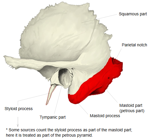
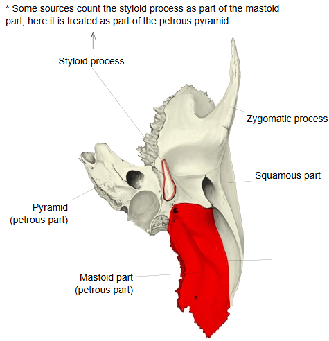
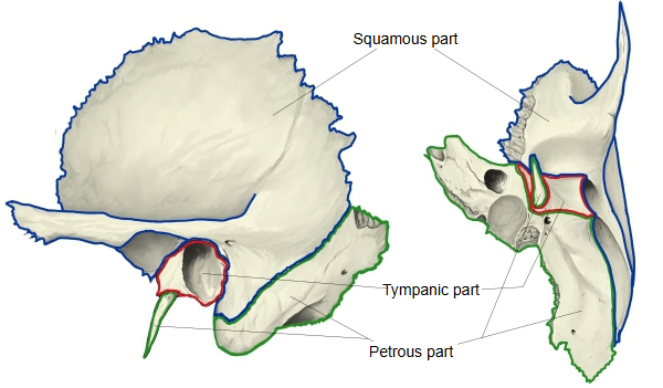
- Squamous part : occupies the middle and the whole upper part of the bone; it has two surfaces, temporal and cerebral.
- Petrous part
- Mastoid part : below the squamous part, from the mastoid process up to the parietal notch.Related terms: mastoid process, mastoid notch, groove for occipital artery, groove for sigmoid sinus, mastoid foramen, occipital border, stylomastoid foramen, canaliculus for chorda tympani, petrotympanic fissure
- Pyramid : a three-sided pyramid running forwards and medially from the boundary of the squamous and mastoid parts, with three surfaces and three borders: the petrous part in the narrow sense.
- Tympanic part : the smallest part: a thin, half-tube-shaped plate that forms the front and lower walls of the external acoustic meatus.
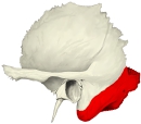 Mastoid part (lateral view)
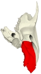 Mastoid part (inferior view)
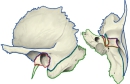 Parts of the temporal bone
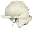 Temporal bone (lateral view), labelled
 Temporal bone (inferior view), labelled
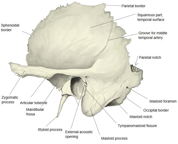
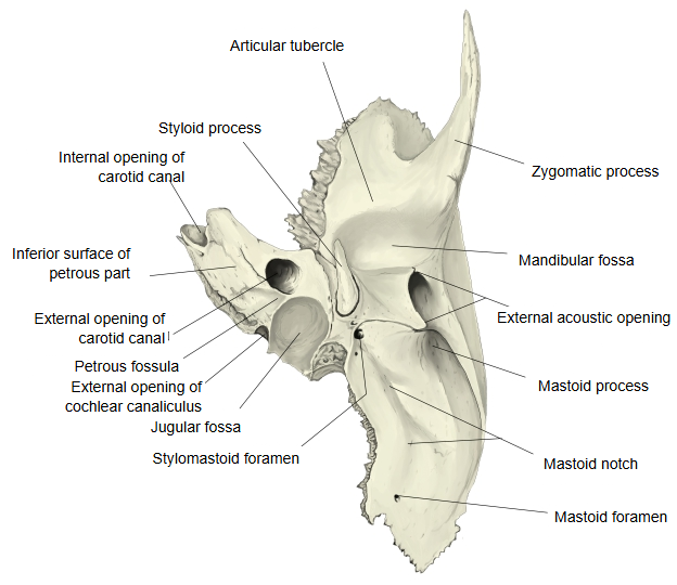
Main features
Lateral surface
1. 1. The lateral surface is rough, because muscles are inserted into it: the sternocleidomastoid, splenius capitis and longissimus capitis.
2. 2. The rounded projection at its front lower part, behind and below the external acoustic opening, is the mastoid process; it lies behind the auricle and is easily felt through the skin.
Like the rest of the lateral surface, it is a site of muscle insertion.
Inferior surface
1. 1. Two grooves run lengthwise, side by side: the medial one is the groove for the occipital artery, which carries the occipital artery, and the lateral one is the mastoid notch, from which the posterior belly of the digastric arises.
2. 2. At the front end of the mastoid notch, behind the styloid process, lies the stylomastoid foramen, the exit of the facial canal and the passage of the facial nerve. Further back, near the posterior border of the mastoid part, lies the mastoid foramen, which carries the mastoid emissary vein to the groove for the sigmoid sinus.
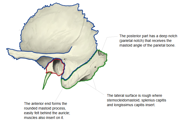
The lateral surface of the mastoid part is rough for muscle attachments; the part that extends downwards behind the external acoustic opening is the mastoid process, into which the sternocleidomastoid is inserted. Behind and medial to the process lies the mastoid notch, the origin of the posterior belly of the digastric, and medial to that the groove for the occipital artery. The inner surface has a deep, long hollow, the groove for the sigmoid sinus, which continues above into the groove for the transverse sinus and below to the jugular foramen. The mastoid foramen near the back border carries the mastoid emissary vein and opens into this groove. The back part of the mastoid part, which joins the occipital squama, is called the occipital border. The facial canal enters the bone at the facial nerve area of the fundus of the internal acoustic meatus, runs forwards and laterally, turns sharply backwards at the geniculum, passes between the tympanic cavity and the semicircular canals, and curves down to the stylomastoid foramen. A little above this foramen the canaliculus for the chorda tympani branches off; the chorda tympani crosses the tympanic cavity between the handle of the malleus and the long limb of the incus and leaves the skull through the petrotympanic fissure.
Reference websites
[Wikipedia]
· Mastoid part of the temporal bone, Wikipedia.
· Mastoid cells, Wikipedia.
[Websites with illustrations]
· The temporal bone (TeachMeAnatomy)
· Temporal bone: anatomy, parts, sutures and foramina (Kenhub)
· Left temporal bone, outer surface (Gray's Anatomy, fig. 137) (Wikimedia Commons)

· Left temporal bone, inferior surface (Gray's Anatomy, fig. 141) (Wikimedia Commons)

Etymology
"Mastoid" comes from the Greek "mastos", breast, and "-eides", like: the mastoid process is shaped like a nipple or breast.
Surfaces
Outer surface: rough; it gives attachment to the occipital belly of the occipitofrontalis and to the posterior auricular muscle, besides the muscles inserted into the mastoid process. It is pierced by many small foramina. The largest, the mastoid foramen, lies near the posterior border; it carries the mastoid emissary vein to the sigmoid sinus and a small branch of the occipital artery to the dura mater. Its size and position vary: it may be absent, or lie in the occipital bone or in the suture between the temporal and occipital bones. Inner surface: it carries the deep, curved groove for the sigmoid sinus, separated from the mastoid air cells only by a thin plate of bone.
Borders
Superior border: broad and serrated; it joins the mastoid angle of the parietal bone, and at its front end the parietal notch receives that angle. Posterior (occipital) border: also serrated; it joins the occipital bone between its lateral angle and the jugular process, at the occipitomastoid suture. In front, the mastoid part is fused above with the descending part of the squamous part; below, it takes part in forming the external acoustic meatus and the tympanic cavity.
Mastoid air cells
A section through the mastoid process shows that it is hollowed out into many spaces, the mastoid air cells, which vary greatly in size and number. In the upper front part of the process they are large, irregular and filled with air; lower down they become smaller, and those at the tip often contain marrow. Sometimes there are no cells and the process is solid. In the upper front part of the bone lies a larger cavity, the mastoid (tympanic) antrum, which communicates with the cells; like them it is filled with air and lined by a continuation of the mucous membrane of the tympanic cavity. Its roof is the thin tegmen tympani, which separates it from the middle cranial fossa; medially the lateral semicircular canal bulges into it, and in front it opens into the epitympanic recess of the tympanic cavity. The antrum is already of fair size at birth; the air cells grow out from it, are well marked by the fifth year and are complete towards puberty.
Development
The mastoid process is absent or very small in the newborn skull. It develops after birth, mainly from about the end of the first year, as the sternocleidomastoid and the other neck muscles develop and pull on the bone.
Related terms
- Mastoid foramen
- Mastoid notch
- Mastoid process
- Mastoid cells
- Petrous part
- Squamous part
- Tympanic part
- Parietal notch
- Groove for sigmoid sinus
References and sources
- Further reading: Mastoid part of the temporal bone, Wikipedia.
- Federative International Programme for Anatomical Terminology (FIPAT). Terminologia Anatomica, 2nd ed. (TA2), 2019.