Synovial joints (Latin: juncturae synoviales; articulationes)
Also called the movable joints
- Overview
- Classification (By shape and movement, By the number of bones, By the number of axes)
- Components
- Lists and illustrations
Overview
Joints can be divided into the immovable joints, which hardly move, and the movable joints, which do move (joints in the narrow sense). The immovable joints are described on the Joints page.
- Immovable joints
- Fibrous joints : syndesmosis, suture, gomphosis
- Cartilaginous joints : e.g. the pubic symphysis
- Bony union : e.g. the fused vertebrae of the adult sacrum
- Movable joints : synovial joints (joints in the narrow sense)
(synovial joints)
- Where two bones meet and move against each other
- A few hundred in the body
- A large part in the hands and feet
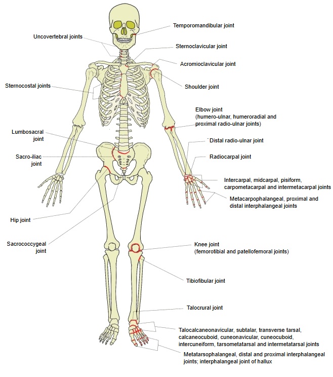
Classification
The movable joints (synovial joints) are classified in three ways:
- By the shape of the articular surfaces and the kind of movement : 1. plane joint 2. ball-and-socket joint 3. cotyloid joint 4. condylar joint 5. hinge joint 6. spiral joint 7. pivot joint 8. saddle joint
- By the number of bones : 1. simple joint 2. compound joint
- By the number of axes of movement : 1. uniaxial joints 2. biaxial joints 3. multiaxial joints
[Classification by the shape of the articular surfaces and the kind of movement]
1. Plane joint (amphiarthrosis): the articular surfaces are flat, or almost flat; movement is limited.
Examples: zygapophysial joints, acromioclavicular joint
2. Ball-and-socket joint (cotyloid joint): the articular head is part of a sphere and the socket is a matching hollow; the joint moves very freely.
Examples: shoulder joint, humeroradial joint, hip joint
3. Condylar joint (ellipsoid joint): the articular head and the socket are oval; the joint moves about the long and the short axis of the oval.
Examples: atlanto-occipital joint, temporomandibular joint, radiocarpal joint
4. Hinge joint: both articular surfaces are parts of a cylinder, like a door hinge; the joint moves in one direction only.
Examples: interphalangeal joints, humeroulnar joint (the main part of the elbow joint)
5. Spiral joint: a variety of the hinge joint in which one articular surface has a ridge and the other a groove, so that the movement follows a slight spiral.
Example: talocrural joint
6. Pivot joint: a uniaxial joint in which the articular head turns like a wheel in the socket.
Examples: superior radioulnar joint, median atlantoaxial joint
7. Saddle joint: both articular surfaces are shaped like a saddle and fit each other at right angles; a biaxial joint.
Examples: carpometacarpal joint of the thumb, sternoclavicular joint
[Classification by the number of bones]
1. Simple joint: a joint formed by two bones.
Examples: shoulder joint, hip joint, interphalangeal joints
2. Compound joint: a joint formed by three or more bones.
Examples: elbow joint, radiocarpal joint
[Classification by the number of axes of movement]
By the number of axes about which they move, the movable joints are divided into three kinds:
(synovial joints)
- Uniaxial joints : move about one axis only (hinge, spiral and pivot joints)
- Biaxial joints : move about two axes at right angles to each other (ellipsoid (= condylar) and saddle joints)
- Multiaxial joints : move about three or more axes (ball-and-socket joints including the cotyloid joint; plane joints including amphiarthroses)
Uniaxial joints (joints that move about one axis only)
| Type | Examples | ||
|---|---|---|---|
| 1 | Hinge joint | Interphalangeal joints, humeroulnar joint | 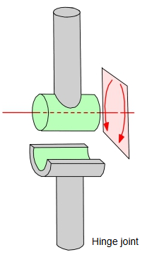 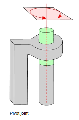 |
| 2 | Spiral joint | Talocrural joint | |
| 3 | Pivot joint | Superior radioulnar joint, median atlantoaxial joint |
Biaxial joints (joints that move about two axes at right angles)
| Type | Examples | ||
|---|---|---|---|
| 1 | Ellipsoid joint | Radiocarpal joint | 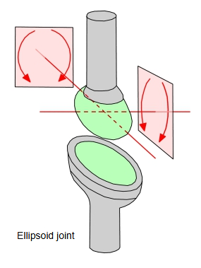 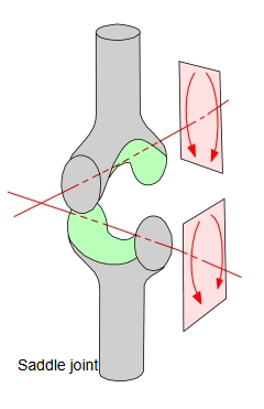 |
| 2 | Condylar joint | Atlanto-occipital joint, temporomandibular joint | |
| 3 | Saddle joint | Carpometacarpal joint of the thumb, sternoclavicular joint |
Multiaxial joints (joints that move about three or more axes)
| Type | Examples | ||
|---|---|---|---|
| 1 | Ball-and-socket joint | Shoulder joint, humeroradial joint | 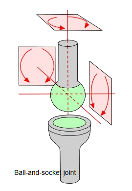 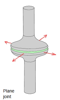 |
| 2 | Plane joint | Zygapophysial joints, acromioclavicular joint | |
| 3 | Amphiarthrosis | Sacroiliac joint | |
| 4 | Cotyloid joint | Hip joint |
Components
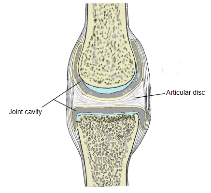
A movable joint is made up of the following parts:
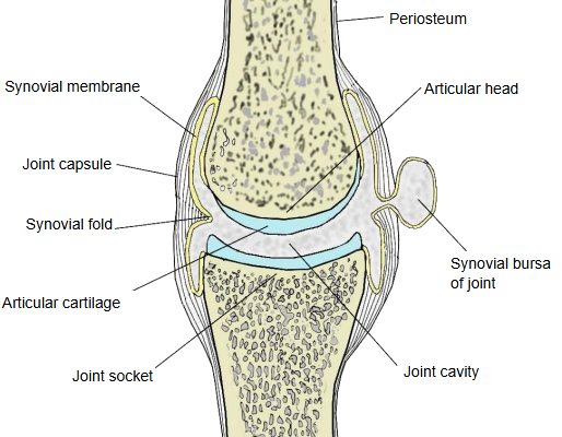
(click to enlarge)
| Name | Notes | |
|---|---|---|
| 1 | Periosteum | The tough fibrous membrane that covers the bone. |
| 2 | Joint cavity | The space between the bones of the joint; it contains synovial fluid. |
| 3 | Synovial bursa of joint | In large joints the synovial membrane, with synovial fluid inside, may bulge out through the joint capsule. |
| 4 | Articular cartilage | The smooth, elastic cartilage that covers the articular surfaces of the bones and acts as a cushion. |
| 5 | Joint socket | The concave end of one of the two bones. |
| 6 | Joint capsule | The connective tissue that continues from the periosteum and encloses the joint. It has two layers: the outer fibrous membrane and the inner synovial membrane. |
| 7 | Synovial fold | A fold of the synovial membrane that projects into the joint cavity. |
| 8 | Synovial membrane | The inner layer of the joint capsule; rich in blood vessels, it secretes the synovial fluid. |
| 9 | Articular head | The convex end of one of the two bones. |
| 10 | Articular disc | A plate of fibrocartilage that divides the joint cavity completely into two. |
| 11 | Synovial fluid | The fluid that fills the joint cavity. |
Lists and illustrations
- List of joints: alphabetical order, by region
- Illustrations of joints
- Joints and ligaments