Latin: scapula · also called the shoulder blade
Overview
- One of the two bones of the shoulder girdle (with the clavicle).
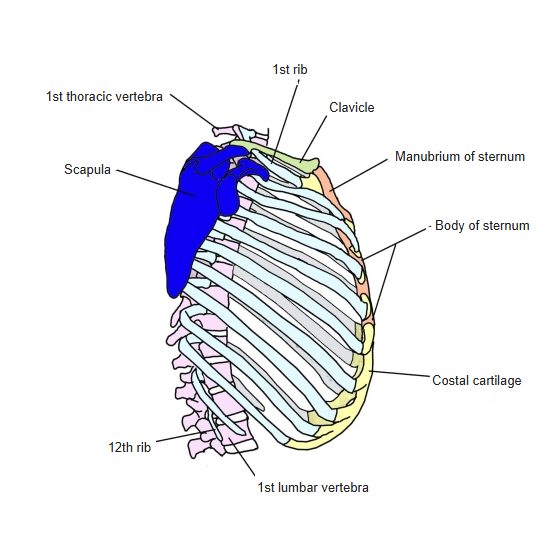
- A flat, triangular bone on the back of the thorax, at the level of the 2nd to 7th (8th) ribs.
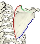
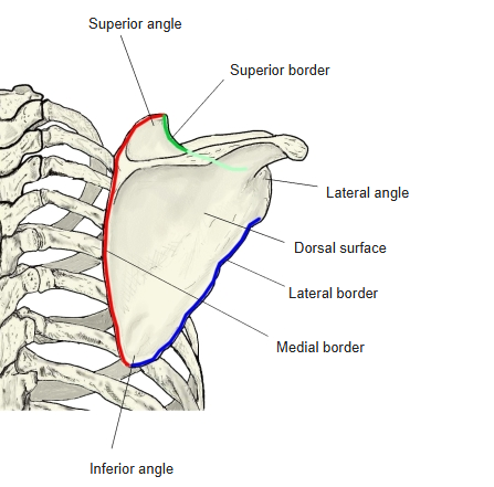
- It has 2 surfaces (costal and dorsal), 3 borders (medial, lateral and superior) and 3 angles (superior, inferior and lateral).
- The spine of the scapula crosses the dorsal surface and ends laterally in the acromion; the coracoid process projects forward from the superior border.
- The glenoid cavity at the lateral angle forms the shoulder joint with the humerus; the scapula therefore has a large influence on movements of the upper limb.
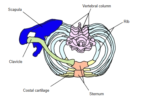
- Medially it is linked to the vertebral column and thorax by many muscles; laterally it meets the clavicle and humerus.
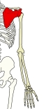
Bones of the upper limb 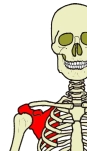
Position (anterior)
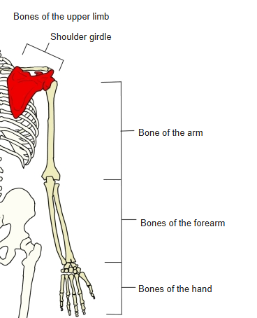
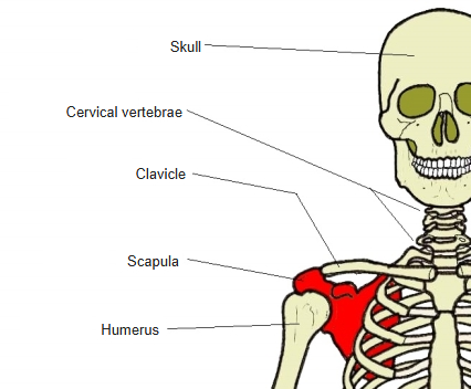
- Bones of the upper limb (one side)
- Shoulder girdle
- Clavicle
- Scapula
- Free upper limb
- Arm
- Humerus
- Forearm
- Radius ← thumb side
- Ulna ← little-finger side
- Hand
- Carpal bones (8)
- Metacarpal bones (5)
- Phalanges (14)
- Arm
- Shoulder girdle
Illustrations
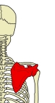
Position (posterior) 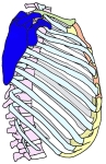
Position (lateral) 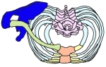
Position (from above)
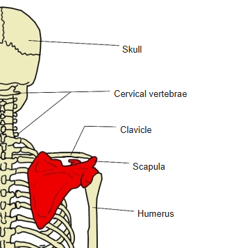
Articulations
| # | Bone | Joint |
|---|---|---|
| 1 | Humerus | Glenohumeral (shoulder) joint |
| 2 | Clavicle | Acromioclavicular joint |
Joints of the scapula
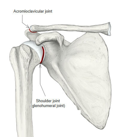
Glenohumeral joint: glenoid cavity ↔ head of humerus. Acromioclavicular joint: clavicular facet of the acromion ↔ acromial facet of the clavicle.
Ligaments
The following ligaments attach to the scapula. The list is intended to be comprehensive but may omit minor ligaments.
| # | Ligament | Connects |
|---|---|---|
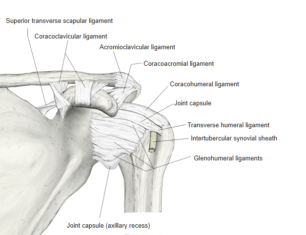
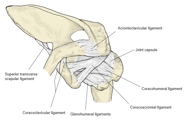
| 1 | Coracohumeral ligament | Coracoid process – greater and lesser tubercles of humerus |
| 2 | Glenohumeral ligaments | Glenoid labrum – anatomical neck of humerus |
| 3 | Acromioclavicular ligament | Acromion – lateral end of clavicle |
| 4 | Coracoclavicular ligament | Coracoid process – underside of the lateral end of clavicle |
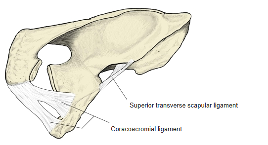
| 5 | Superior transverse scapular ligament | Bridges the suprascapular notch |
| 6 | Inferior transverse scapular ligament | Spans the spinoglenoid notch at the back of the neck of the scapula |
| 7 | Coracoacromial ligament | Coracoid process – acromion |
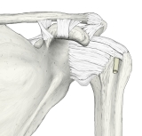
Right shoulder joint (anterior) 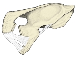
Ligaments of the scapula 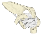
Right shoulder joint (from above)
Muscle attachments
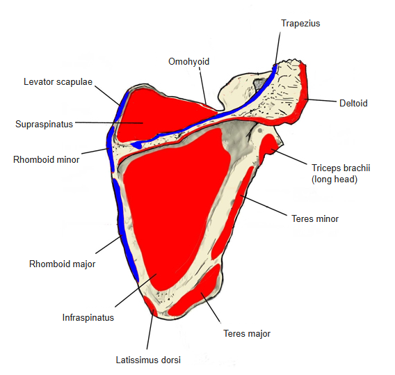
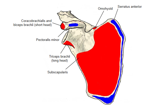
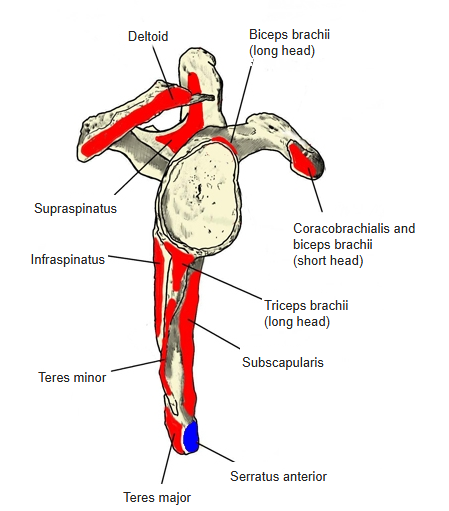
- Origin of: Supraspinatus, Infraspinatus, Subscapularis, Teres minor, Teres major, Deltoid, Omohyoid, Latissimus dorsi, Triceps brachii, Biceps brachii, Coracobrachialis
- Insertion of: Trapezius, Levator scapulae, Rhomboid major, Rhomboid minor, Serratus anterior, Pectoralis minor
Origins: supraspinatus — supraspinous fossa; infraspinatus — infraspinous fossa; subscapularis — subscapular fossa; teres minor — dorsal surface near the lateral border; teres major — dorsal surface of the inferior angle and the lower part of the lateral border; deltoid — spine of scapula; omohyoid — superior border; latissimus dorsi — inferior angle (some fibres); triceps brachii (long head) — infraglenoid tubercle; biceps brachii — long head from the supraglenoid tubercle, short head from the coracoid process; coracobrachialis — coracoid process. Insertions: trapezius — acromion and spine of scapula; levator scapulae — medial border, upper third; rhomboid major and minor — medial border; serratus anterior — medial border and inferior angle; pectoralis minor — coracoid process.
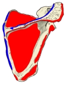
Muscle attachments (posterior) 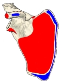
Muscle attachments (anterior) 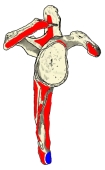
Muscle attachments (lateral)
Surfaces, borders and angles
- Surfaces: the costal (anterior) surface faces the thorax; the dorsal (posterior) surface faces away from the thorax.
- Borders: medial border, lateral border and superior border.
- Angles: superior angle, inferior angle and lateral angle.
Borders and angles
Anomalies
- In many non-mammalian vertebrates the coracoid is a separate bone; in humans the coracoid process rarely remains separate.
- The subscapular fossa may be deeply hollowed (so-called scaphoid scapula).
- The suprascapular notch is often very shallow; the superior transverse scapular ligament bridging it may ossify and turn the notch into a foramen. A small foramen is rarely found near a shallow notch.
Etymology
- In Latin, scapulae (plural) meant the shoulders or shoulder blades, and also the back.
- In the 17th century the French anatomist Jean Riolan first used scapula for this bone, linking it to the Greek skaptein (“to dig”) because the bone resembles a shovel.
References and sources
- Further reading: Scapula, Wikipedia.