Latin: maxilla
Overview
- The paired upper jaw bone. The two maxillae meet in the midline and form most of the upper jaw, the floor of the orbit, the lateral wall and floor of the nasal cavity and most of the hard palate; they carry the upper teeth.
- Each maxilla has a body, containing the large maxillary sinus, and four processes: frontal, zygomatic, alveolar and palatine.
- It meets more bones than any other facial bone, and many muscles of facial expression arise from its front surface.
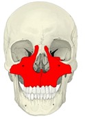
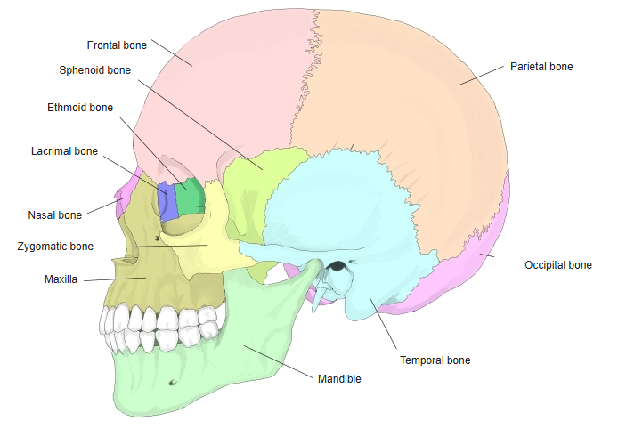
Skull (anterior) 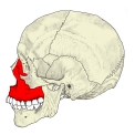
Skull (left lateral)
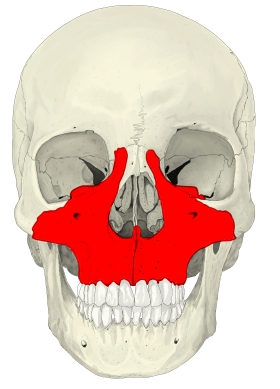
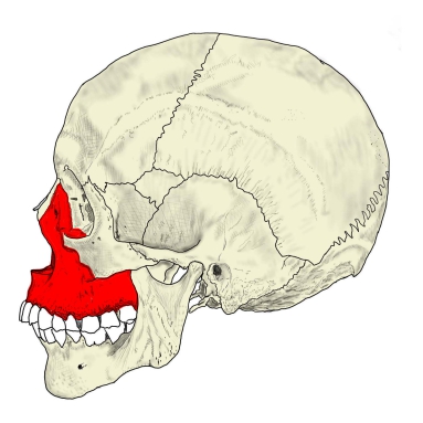
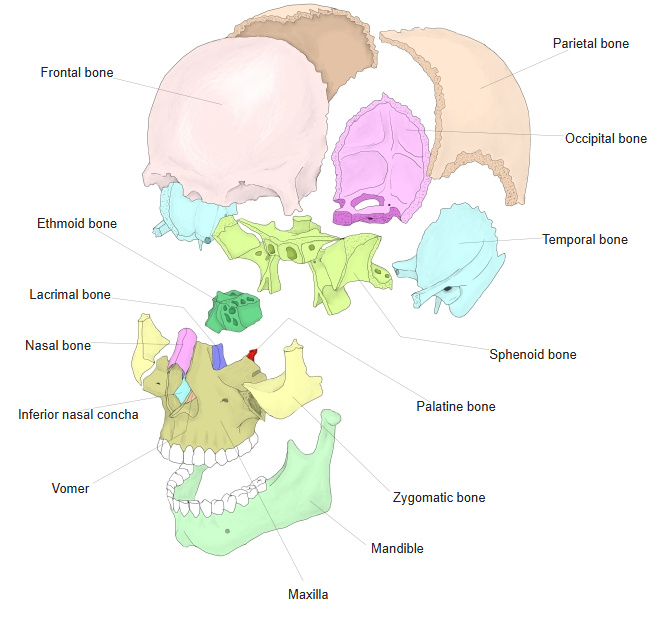
- Bones of the skull
- Neurocranium (cranial bones)
- Frontal bone
- Parietal bone (2)
- Occipital bone
- Temporal bone (2)
- Sphenoid bone
- Ethmoid bone
- Viscerocranium (facial bones)
- Nasal bone (2)
- Vomer
- Lacrimal bone (2)
- Inferior nasal concha (2)
- Maxilla (2)
- Zygomatic bone (2)
- Palatine bone (2)
- Mandible
- Hyoid bone
- Neurocranium (cranial bones)
Illustrations
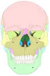
Bones of the skull (anterior) 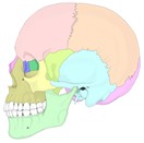
Bones of the skull (lateral) 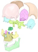
Bones of the skull (separated)
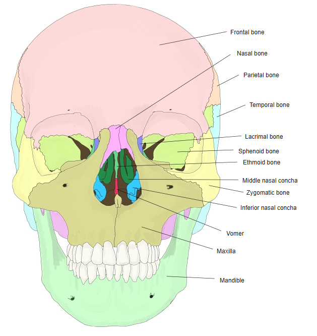
Articulations
| # | Bone | Joint |
|---|---|---|
| 1 | Frontal bone | Frontal process |
| 2 | Ethmoid bone | Orbital surface and ethmoidal crest |
| 3 | Nasal bone | Frontal process (nasomaxillary suture) |
| 4 | Lacrimal bone | Lacrimal border of the frontal process |
| 5 | Zygomatic bone | Zygomatic process |
| 6 | Inferior nasal concha | Conchal crest and maxillary hiatus |
| 7 | Palatine bone | Nasal surface and palatine process (transverse palatine suture) |
| 8 | Vomer | Nasal crest |
| 9 | Opposite maxilla | Intermaxillary and median palatine sutures |
| 10 | Sphenoid bone | Sometimes, at the orbital surface |
Muscle attachments
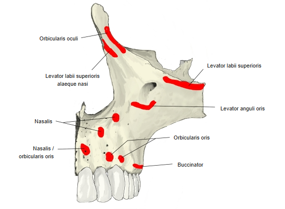
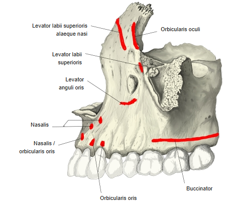
- Origin of: Orbicularis oculi, Levator labii superioris alaeque nasi, Levator labii superioris, Levator anguli oris, Nasalis, Orbicularis oris, Buccinator, Medial pterygoid (superficial head, from the maxillary tuberosity), Masseter (in part, from the zygomatic process)
- Insertion of: none
Most of these are muscles of facial expression arising from the front of the body and the alveolar process; buccinator arises from the alveolar process opposite the molars.
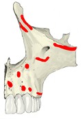
Muscle attachments (anterior) 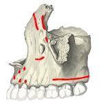
Muscle attachments (anterolateral)
Body
- Anterior surface – the infraorbital foramen opens below the orbital margin; below it lies the canine fossa. Medially it ends at the nasal notch and the anterior nasal spine.
- Infratemporal surface – faces backwards; its lower part bulges as the maxillary tuberosity and carries the small alveolar foramina that lead into the alveolar canals.
- Orbital surface – forms most of the floor of the orbit; the infraorbital groove runs forwards on it and becomes the infraorbital canal.
- Nasal surface – forms part of the lateral wall of the nasal cavity, with the large maxillary hiatus opening into the sinus, the groove for the lacrimal sac in front and the conchal crest for the inferior nasal concha.
- Maxillary sinus – the largest paranasal sinus, filling the body and opening into the middle nasal meatus.
Processes
- Frontal process – rises between the nasal and lacrimal bones to meet the frontal bone; it carries the anterior lacrimal crest and the ethmoidal crest.
- Zygomatic process – a rough projection that joins the zygomatic bone; the zygomaticoalveolar crest runs down from it to the first molar.
- Alveolar process – the curved alveolar arch carrying the sockets of the upper teeth, separated by interalveolar septa; the roots raise the alveolar yokes on its outer surface.
- Palatine process – a horizontal plate forming the front three-quarters of the hard palate. The two processes meet at the median palatine suture, rising as the nasal crest above; the incisive fossa and incisive canals lie at the front, and the transverse palatine suture joins the palatine bones behind.
References and sources
- Further reading: Maxilla, Wikipedia.