Optic canal (Latin: canalis opticus)
Also called Optic foramen (often used for its opening, or for the whole canal)
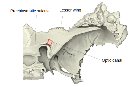
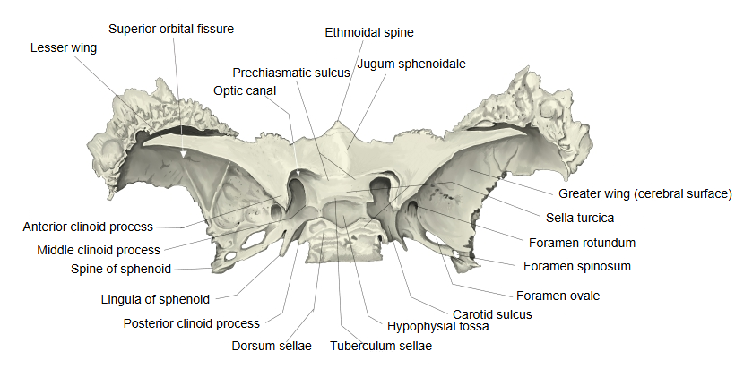
· The lesser wing of the sphenoid bone arises from the body by two roots, an upper (anterior) root and a lower (posterior) root, and the optic canal lies between these two roots and the side of the body. The lower root (optic strut) separates it from the superior orbital fissure.
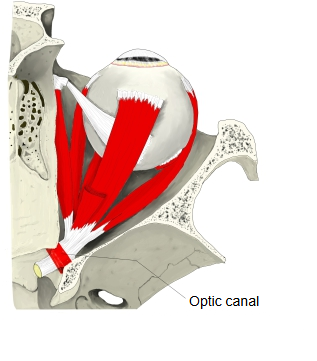
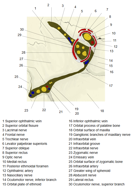
The canal runs forwards and laterally; the two canals are about 25 mm apart at their cranial ends and about 30 mm apart at their orbital ends. The optic nerve passes through it with its meningeal sheaths, and the ophthalmic artery lies below and lateral to the nerve, with some sympathetic fibres. Sources differ on whether the canal is narrowest at the front or in the middle. Because the canal is tight, fractures or swelling here can compress the optic nerve and cause loss of vision (traumatic optic neuropathy).
the
sphenoid
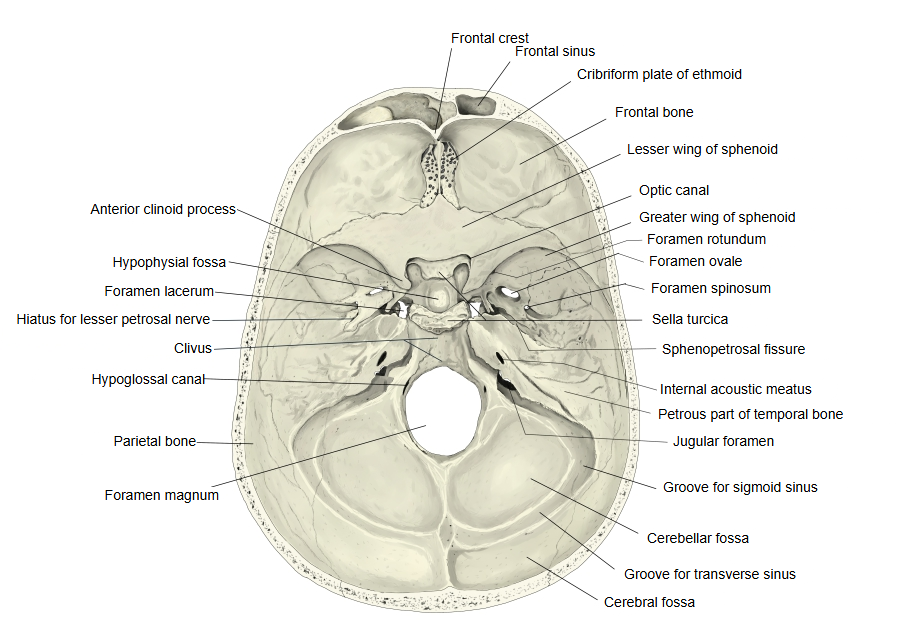
- Upper surface : faces the cranial cavity and forms the middle part of the cranial fossae. Joins the ethmoid bone in front.
Features: sella turcica, hypophysial fossa, tuberculum sellae, dorsum sellae, middle clinoid process, posterior clinoid process, prechiasmatic sulcus, carotid sulcus, sphenoidal lingula. - Lower surface : forms part of the roof of the nasal cavity and the pharynx (nasopharynx). Joins the vomer.
- Anterior surface : faces the nasal cavity. Joins the ethmoid bone.
Features: sphenoidal crest, sphenoidal rostrum, sphenoidal concha (aperture of the sphenoidal sinus). - Posterior surface : a four-sided rough surface. Joins the occipital bone (spheno-occipital synchondrosis).
- Lateral surfaces : the greater wing and the pterygoid process arise from each side.
· = optic foramen
Although it is a canal, it is short (only about 1 cm), so many sources treat it as a foramen; they then write optic canal = optic foramen.
Other sources, such as Wikipedia, keep the two names apart.
In that usage the optic foramen is the opening of the optic canal (some texts mean only its orbital opening).
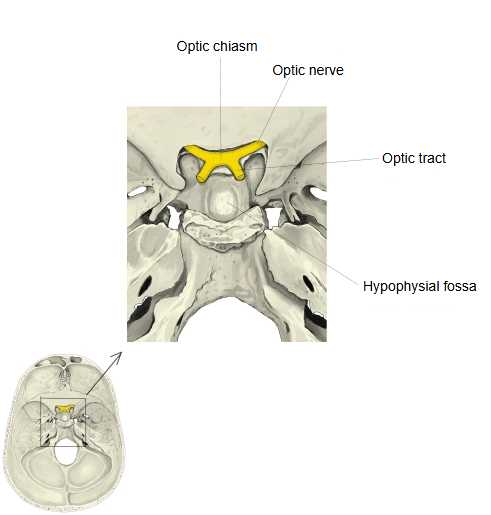
· The prechiasmatic sulcus (chiasmatic groove) runs transversely in front of the tuberculum sellae, and at each end it continues into the optic canal. It is named after the optic chiasm, although the chiasm usually lies above and behind it rather than in the groove.
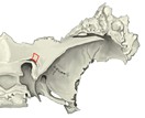 Sphenoid bone (lesser wing, oblique view)
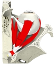 Right orbit (section from above)
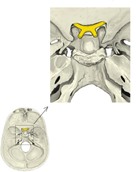 Optic chiasm on the cranial base
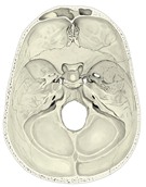 Cranial base (internal surface)
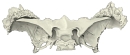 Sphenoid bone (superior)
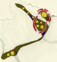 Openings at the apex of the right orbit
Reference websites
[Wikipedia]
· Optic canal, Wikipedia.
· Optic nerve, Wikipedia.
[Websites with illustrations]
· Optic canal (Kenhub)
· Sphenoid bone, superior and posterior views, with the optic canal (Health Jade)
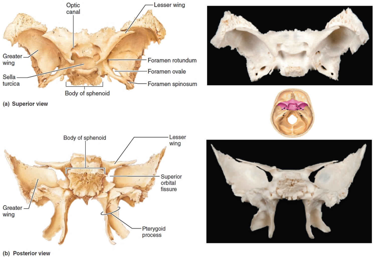
· Bony landmarks of the sphenoid wings and pterygoid process (TeachMeAnatomy)

Where it is found
Related terms
References and sources
- Further reading: Optic canal, Wikipedia.
- Further reading: Optic nerve, Wikipedia.
- Federative International Programme for Anatomical Terminology (FIPAT). Terminologia Anatomica, 2nd ed. (TA2), 2019.