Lymph node (Latin: nodus lymphoideus)
Also called the lymph gland (older term)
- Overview
- Lymph node and lymphoid nodule
- Structure (Capsule, Cortex, Paracortex, Medulla, Lymph sinuses)
- Cells of the lymph node
- Functions
- Distribution of reticular fibres
- Nerves and blood vessels
- Reference websites
- (Wikipedia, websites with illustrations)
- Where it is found
- Related terms
Overview
lymphatic
system
- Lymph (fluid)
- Lymphatic vessels
- Lymphatic capillaries : where tissue fluid first seeps in; diameter about 10–60 µm.
- Small and medium lymphatic vessels : carry the lymph from the capillaries; diameter about 100–500 µm.
- Lymphatic trunks : receive the lymph from the small and medium vessels; diameter about 0.2–2 mm.
- Thoracic duct and right lymphatic duct : the main trunks that open directly into the veins; the thoracic duct collects about three quarters of the body's lymph.
- Lymph nodes : bean-shaped, about 2–30 mm across, roughly 600–800 in number. Function: filtering the lymph and multiplying T and B cells.
- Lymphoid tissue
- Primary lymphoid organs : thymus, bone marrow.
- Secondary lymphoid organs : lymph nodes, spleen, mucosa-associated lymphoid tissue.
- Diffuse lymphoid tissue : lymphocytes scattered in the lamina propria of the mucosa and elsewhere.
· Small lymphatic vessels tend to carry small lymph nodes and large vessels large ones, but this is not true in every case.
1. 1. The size changes with immune activity and health.
With inflammation or infection a lymph node swells and enlarges, so even at the same site its size varies with the state of health. Most normal nodes are less than 1 cm long.
2. 2. Differences between regions
Some regions, such as the axilla, the groin and the neck, have many nodes that are large to begin with.
· Lymph nodes are found all over the body; regions with especially many are:
1. 1. Neck (cervical nodes): about 200
2. 2. Axilla (armpit): about 20–30
3. 3. Groin (inguinal nodes, at the root of the thigh): about 10–20
4. 4. Many more in the chest (mediastinum), the abdomen (along the large vessels) and the pelvis
Lymph node and lymphoid nodule
"Lymph node" and "lymphoid nodule" sound alike, but they are quite different things.
1. 1. A lymphoid nodule has no capsule around it, unlike a lymph node.
2. 2. A lymph node is an organ placed on the lymphatic vessels, while a lymphoid nodule lies independently of them (in the mucosa of the gut, airways and other tracts, singly or in groups such as the tonsils and Peyer's patches).
| Feature | Lymph node | Lymphoid nodule | |
|---|---|---|---|
| 1 | Form | An oval organ with a capsule, a cortex and a medulla | A small follicle-like cluster of lymphocytes; no capsule |
| 2 | Make-up | B-cell follicles + T-cell zone + macrophages | Mainly a B-cell follicle, often with a germinal centre |
| 3 | Position | Along the lymphatic vessels throughout the body | Inside lymph nodes, or scattered in the mucosa and submucosa |
| 4 | Function | Filters the lymph of the whole body; starts immune responses | Local immune responses (antigen recognition, antibody production) |
| 5 | Connection | Linked to afferent and efferent lymphatic vessels | Local; lymph flow through it is limited |
Structure
A lymph node can be divided into the capsule, its outermost layer, and the inner structure, which is divided further into several parts.
of a
lymph node
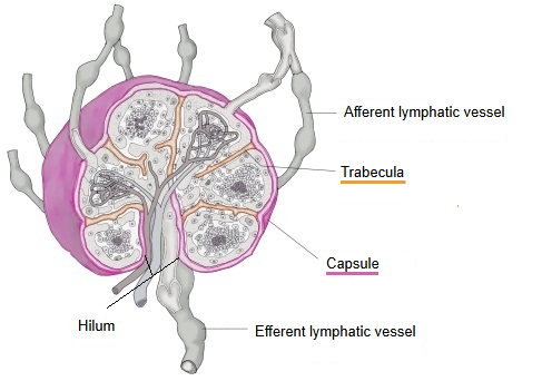
- Capsule : the outermost layer of connective tissue that wraps the node; it sends fibrous septa (trabeculae) inward.
- Inside
- Cortex
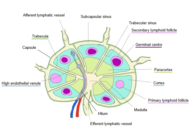
- Follicles : spherical groups of B cells in the outer part; they form germinal centres.
- Parafollicular area : the area between the follicles, where T cells gather.
- Paracortex : a region typical of lymph nodes between the cortex and the medulla; mainly a dense T-cell zone.
- Medulla
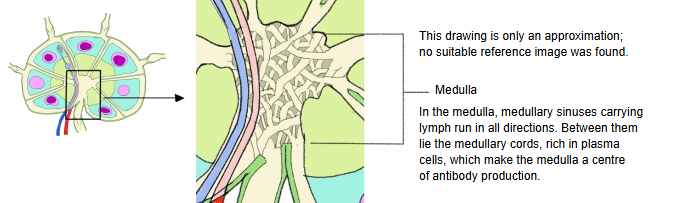
- Medullary cords : plasma cells, B cells, macrophages and others; function: the centre of antibody production.
- Medullary sinuses : spaces through which lymph flows; macrophages catch foreign matter and debris.
- Lymph sinuses : the channels of the lymph, lined by endothelial cells: subcapsular (marginal) sinus, trabecular (intermediate) sinuses and medullary sinuses.
- Cortex
[Capsule]
A thick, tough layer of fibrous connective tissue (collagen fibres) that wraps the whole lymph node.
· The fibre bundles of the capsule may also contain some elastic fibres.
Function: it keeps the shape of the lymph node and supports the cortex and medulla inside.
· Afferent lymphatic vessels enter the lymph node and efferent lymphatic vessels leave it. Node and vessels may look alike from outside, but the node is wrapped in a connective tissue capsule, while the wall of a lymphatic vessel is built on a lining of endothelial cells.
Parts that continue inward
· Blood vessels branch from the capsule and reach the follicles of the cortex, the paracortex and the medullary cords.
· Nerves (sympathetic nerve fibres) also run inward along the vessels and influence the blood flow and the activity of the immune cells.
[Cortex]
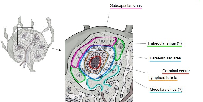
The cortex is the outer part of the lymph node, just inside the capsule. It can be divided into the follicles and the parafollicular area.
Follicles
A follicle is a cluster of cells, mainly B cells, in the cortex. There are two kinds:
Primary lymphoid follicle: made mostly of resting (naive) B cells that have not been activated.
Secondary lymphoid follicle: a germinal centre appears, where activated B cells multiply and differentiate into centroblasts (dark zone) and centrocytes (light zone).
· The lymph node is divided into several compartments by the trabeculae that run in from the capsule. Not every compartment has a follicle: some have none and some have several.
· In a healthy lymph node there are many primary follicles, and secondary follicles form in some places in response to antigens, so the cortex usually contains both kinds.
Parafollicular area
The parafollicular area is the T-cell zone around the follicles; many dendritic cells are also seen there.
The proportions in the table below are rough estimates; sources give different figures.
| Cell | Share (approx.) | Comment | |
|---|---|---|---|
| 1 | T cells (CD4⁺/CD8⁺) | about 60–70% | Mainly CD4⁺ helper T cells. Activated by antigen presentation, they activate B cells and other T cells. CD8⁺ cells are fewer. |
| 2 | Dendritic cells | about 10–15% | Present antigens to T cells; important for antigen recognition in the node. |
| 3 | Macrophages | about 5–10% | Engulf antigens and remove debris; also help to activate T cells. |
| 4 | B cells | about 5–10% | Naive B cells and a few memory B cells; mostly in and around the follicles, few in the parafollicular area. |
| 5 | Others | <5% | NK cells, immature cells and others; varies with the situation. |
Function
1. 1. Gathering and activating T cells
Naive T cells are activated by antigen presentation from dendritic cells.
2. 2. The starting point of the immune response
Activated naive T cells develop into the following cells and begin their immune work.
· Helper T cells ⇒ stay in the node and help B cells to produce antibodies.
· Killer T cells ⇒ leave with the lymph, reach the whole body and destroy infected cells.
3. 3. A place where cells interact
Contact between T cells and dendritic cells
Interaction between T cells and B cells
[Paracortex]
· Besides T cells, dendritic cells and others are found there.
· High endothelial venules (venules lined by tall, plump endothelial cells) are seen there.
· Some sources call this the part with the most blood vessels and nerves, while others give the medulla.
Function
1. 1. The T-cell zone
T cells are activated (multiply and differentiate) by antigen presentation from dendritic cells.
2. 2. The gateway through which lymphocytes enter the node from the blood
Naive T cells leave the blood through the high endothelial venules (HEVs), enter the paracortex and wait there.
3. 3. Closely involved in the swelling of lymph nodes (especially in viral infections)
In a viral infection the T cells multiply, the paracortex enlarges markedly, and the whole node swells.
[Medulla]
Medullary sinuses
· The kidney, the suprarenal gland, the brain and other organs also have a "medulla", but the medullary sinus is a name used only for the lymph node.
· The medullary sinuses take up most of the medulla.
Textbooks do not usually give the proportion of sinuses to cords; a rough estimate is about 60–75% sinuses.
· Some sources call this the part with the most blood vessels and nerves, while others give the paracortex.
Lymph flow in a lymph node
Lymph from every compartment drains into the efferent vessel.
Medullary cords
The cells in the medullary cords are:
| Cell type | Notes | |
|---|---|---|
| 1 | Plasma cells | The most numerous cells and the main cells of the cords; they stand out as antibody-producing cells and may make up more than half of the cells. |
| 2 | B cells | Include B cells activated in the germinal centres and memory B cells; fewer than plasma cells. |
| 3 | Macrophages | Mostly where the cords meet the medullary sinuses, catching antigens in the lymph; few inside the cords. |
| 4 | Dendritic cells | Few; they present antigens, at the ends of the cords and near the sinuses. |
| 5 | T cells | Not many in the cords; most are in the paracortex, but some are found here. |
[Lymph sinuses]
of lymph
sinus
- Subcapsular sinus : = marginal sinus; just under the capsule; lymph from the afferent lymphatic vessels passes here first.
- Trabecular sinus : = cortical (intermediate) sinus; runs along the trabeculae (connective tissue septa).
- Medullary sinus : in the medulla; drains finally into the efferent lymphatic vessel.
Note: other names, such as "intermediate sinus" and "cortical sinus", are also used; they usually mean the same channels as the trabecular sinuses.
Lymph flow in a lymph node
Lymph from every compartment drains into the efferent vessel.
Subcapsular sinus
· The place where the lymph from the afferent lymphatic vessels first enters the node; it covers almost the whole node.
(an almost continuous space all the way round)
· Also called the subcapsular sinus or marginal sinus ("subcortical sinus" in some texts).
· Macrophages are placed there and catch and present antigens.
The subcapsular sinus is where anything suspicious entering the node is held first.
Trabecular sinus
· A lymph channel continuing from the subcapsular sinus; it runs along the trabeculae (connective tissue septa) through the cortex toward the medulla.
· Also called the cortical or intermediate sinus.
· Its lumen is fairly wide, and its inner surface is lined by sinus endothelium (a discontinuous endothelium).
· Reticular fibres (reticulin fibres) stretch across the lumen of the sinus.
· The trabecular sinus acts as a passage that guides the lymph (and the cells in it).
Medullary sinus
· The lymph channels in the medulla of the node: the last passage before the lymph leaves the node through the efferent lymphatic vessels.
(the boundary between the medullary sinuses and the efferent lymphatic vessel is not sharp; one passes gradually into the other)
Medullary sinus: discontinuous sinus endothelium ⇒ efferent lymphatic vessel: a more continuous endothelium
· A branched, tube-like system of spaces, often running along the trabeculae.
· Macrophages (and also plasma cells and a few lymphocytes) are present; antigens are caught here and antibodies are produced nearby.
Cells of the lymph node
A short list of the cells found in a lymph node.
| Cell | Main location | Function | |
|---|---|---|---|
| 1 | B cells | Cortical follicles (primary and secondary) | Prepare for antibody production, form follicles, differentiate into plasma cells and memory B cells |
| 2 | T cells | Paracortex | Recognize antigens, direct cell-mediated immunity, regulate the immune response |
| 3 | Plasma cells | Medullary cords | Produce antibodies |
| 4 | Dendritic cells | Paracortex, follicles | Present antigens; activate B cells and T cells |
| 5 | Macrophages | Medullary cords, medullary sinuses, follicles | Catch antigens and dead cells; clean the lymph |
| 6 | Reticular cells | Throughout the node (cortex, paracortex, medullary cords, trabeculae) | Make reticular fibres; support lymphocytes and other immune cells; keep the structure |
| 7 | Endothelial cells | High endothelial venules (HEVs), capillaries | Form the vessel walls; control the passage of lymphocytes |
| 8 | Smooth muscle-like cells (myofibroblast-like cells) | Trabeculae, capsule | Contraction of the trabeculae and capsule; support of the vessels |
Nerves: nerve fibres are present, but there are no nerve cell bodies in a lymph node.
Functions
1. 1. Filtering the lymph
Foreign matter in the lymph (bacteria, viruses, debris) is caught.
· A mesh of fine fibres stretched inside the node acts as the filter.
· (immune reaction) Macrophages engulf what is caught (phagocytosis).
2. 2. A site of immune responses
Immune cells such as B cells, T cells and macrophages are activated here; activated B cells become plasma cells, and effector and memory lymphocytes leave with the lymph and return to the blood.
3. 3. Adjusting the flow of body fluid
Part of the route that returns surplus extracellular fluid, as lymph, to the blood.
Distribution of reticular fibres
The reticular fibres of a lymph node (secreted by reticular cells) form the framework that keeps the shape of the node.
The table lists how the reticular fibres are distributed in each part.
| Part | Features | |
|---|---|---|
| 1 | Capsule | Thick outer connective tissue; few reticular fibres, present as fibre bundles |
| 2 | Cortex | Form the framework that supports the B cells in the primary and secondary follicles |
| 3 | Paracortex | In this zone of T cells the reticular fibres spread as a mesh that supports the T cells |
| 4 | Medulla | Present in the medullary cords, supporting plasma cells and lymphocytes |
| 5 | Lymph sinuses | Few reticular fibres; the walls are formed mainly by endothelium, and some fibres cross the sinuses as a supporting framework |
Nerves and blood vessels
Supplying the capsule
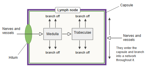
Vessels and nerves around the lymph node supply the capsule directly. After entering the capsule, they form a network that covers the whole capsule.
· Vessels: small arteries and veins; at their ends they form a capillary network.
· Nerves: sensory and sympathetic fibres, mainly running with the arteries.
Supplying the inside
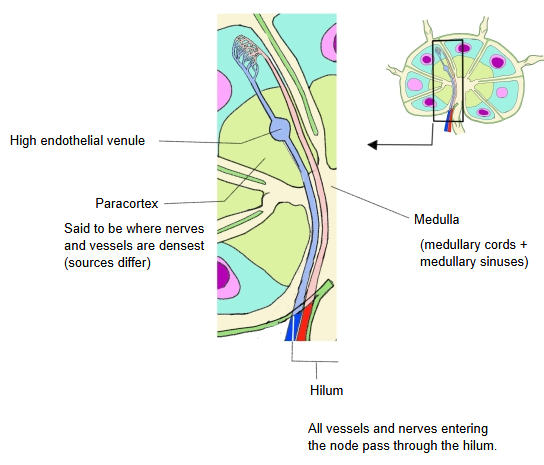
· Vessels: the vessels entering at the hilum run through the medulla (in the medullary cords, not in the sinuses) and then into the trabeculae (in their connective tissue) toward the outside.
On their way through the medulla and the trabeculae they give off side branches.
The venules include the high endothelial venules (lined by cuboidal to tall endothelial cells), typical of lymphoid organs.
· Nerves: almost all sympathetic; they run along the vessels and spread widely inside the node.
Sources differ on which region is most densely supplied with vessels and nerves: some give the paracortex, others the medulla. The table follows the second view.
| Region | Density | Features | |
|---|---|---|---|
| 1 | Medullary cords | highest | Many plasma cells gather here, and dense capillaries supply the oxygen and nutrients needed for antibody production. Nerves also run along the vessels. |
| 2 | Paracortex | medium | The T-cell zone. Its vessels serve mainly the passage of lymphocytes through the HEVs (high endothelial venules); vessels and nerves are less dense than in the medullary cords. |
| 3 | Follicles | low | The B-cell zone. Few vessels and nerves; they mainly supply oxygen and nutrients when germinal centres form. |
Reference websites
[Wikipedia]
· Lymph node, Wikipedia.
[Websites with illustrations]
· Schematic of a lymph node showing the lymph sinuses (Wikimedia Commons)

· Lymph node structure (Wikimedia Commons)

· Lymph nodes (SEER Training (NCI))
Where it is found
Related terms
References and sources
- Further reading: Lymph node, Wikipedia.
- Federative International Programme for Anatomical Terminology (FIPAT). Terminologia Anatomica, 2nd ed. (TA2), 2019.