Pterygoid process (Latin: processus pterygoideus)
Also called the pterygoid process of the sphenoid
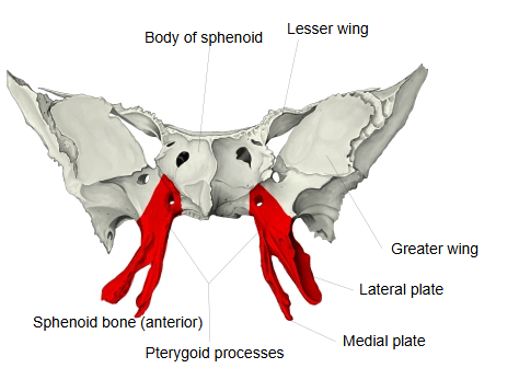
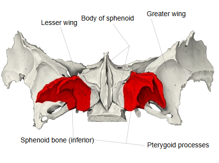
- Body : the central part of the sphenoid; it contains the sphenoidal sinus. Its surfaces are the superior, anterior, inferior and posterior surfaces and two lateral surfaces.
- Processes
- Greater wing : the largest process; it forms part of the middle cranial fossa and of the side wall of the skull.
- Lesser wing : projects to the left and right from the front of the body; it forms part of the anterior cranial fossa.
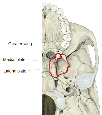
- Pterygoid process : projects downwards from the body and the greater wing; it consists of two plates, the medial plate and the lateral plate.
- Medial plate : the superior constrictor of the pharynx arises from the lower part of its posterior border and from the hamulus.Related terms: pterygoid hamulus, groove of pterygoid hamulus, scaphoid fossa, vaginal process, vomerovaginal canal / groove, palatovaginal canal / groove
- Lateral plate : shorter and broader than the medial plate; the lower head of the lateral pterygoid arises from its outer surface.Related terms: pterygospinous process
- Both plates : related terms common to the two plates: pterygoid notch, pterygoid fossa, pterygoid canal.
Other features
The main features of the pterygoid process are summarised below.
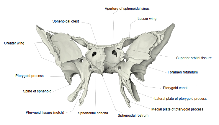
· The plate-like process that runs medially from the root of the medial plate is the vaginal process.
· The vaginal process lies against the ala of the vomer and, with the under surface of the sphenoid bone, forms the vomerovaginal canal.
· The canal running from front to back on the under side of the vaginal process is the palatovaginal canal; it is often only a groove (palatovaginal groove).
(Whether canal or groove, it carries the pharyngeal branch of the maxillary artery and the pharyngeal nerve from the pterygopalatine ganglion.)
· The deep hollow on the back of the pterygoid process, between the medial plate and the lateral plate, is the pterygoid fossa; most of the medial pterygoid muscle arises from it.
· Above and medial to the pterygoid fossa is a shallow oval depression, the scaphoid fossa, from which the tensor veli palatini arises.
· The canal that runs from front to back through the root of the pterygoid process is the pterygoid canal; it carries the nerve of the pterygoid canal and the artery and vein of the pterygoid canal.
· The pterygoid canal opens in front into the pterygopalatine fossa. Below this opening, the front of the process carries a groove running downwards, the greater palatine groove, which joins the maxilla and the palatine bone to form the greater palatine canal.
· The two plates are joined in front. Below, they are separated by the pterygoid fissure (notch), into which the pyramidal process of the palatine bone fits.
· The front surface of the process, with the maxillary surface of the greater wing, forms the back wall of the pterygopalatine fossa.
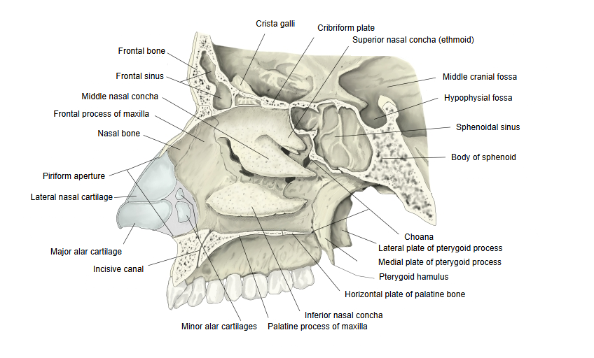
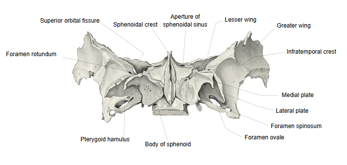
· The medial plate is narrow and long and forms part of the lateral wall of the back of the nasal cavity, beside the choana. Its lower end is a small hook, the pterygoid hamulus, around which the tendon of tensor veli palatini turns. Its upper part carries the groove for the auditory tube. The superior constrictor of the pharynx arises from the lower part of its posterior border and from the hamulus, to which the pterygomandibular raphe is also attached.
· The lateral plate is shorter but broader. Its outer surface gives origin to the lower head of the lateral pterygoid; its inner surface, in the pterygoid fossa, to most of the medial pterygoid. Its back edge may bear a pterygospinous process, joined to the spine of the sphenoid by the pterygospinous ligament.
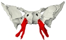 Sphenoid bone (anterior view)
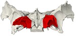 Sphenoid bone (inferior view)
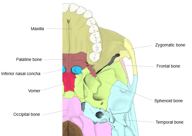
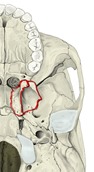 External cranial base
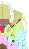 External cranial base (bones colour-coded)
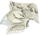 Right nasal cavity (lateral wall)
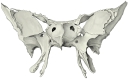 Sphenoid bone (anterior view)
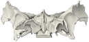 Sphenoid bone (inferior view)
Reference websites
[Wikipedia]
· Pterygoid processes of the sphenoid, Wikipedia.
· Sphenoid bone, Wikipedia.
[Websites with illustrations]
· Sphenoid bone: anatomy, function and development (Kenhub)
· Sphenoid bone, anterior and inferior surfaces (Gray's Anatomy, fig. 146) (Wikimedia Commons)

· Sphenoid bone, upper and posterior surfaces (Gray's Anatomy, fig. 147) (Wikimedia Commons)

Where it is found
Related terms
References and sources
- Further reading: Pterygoid processes of the sphenoid, Wikipedia.
- Federative International Programme for Anatomical Terminology (FIPAT). Terminologia Anatomica, 2nd ed. (TA2), 2019.