Anal canal (Latin: canalis analis)
- Overview
- Divisions (upper anal canal, intermediate zone, lower anal canal)
- Structure
- Vessels and nerves (arteries, veins, lymphatics, nerves)
- Related terms
- References
Overview
· Some anatomy textbooks count the anal canal as part of the rectum.
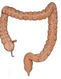
large intestine
- Cecum : length 6–9 cm; the beginning of the large intestine; temporarily stores the contents from the small intestine.
- Vermiform appendix : a tube about 6–10 cm long; helps to keep the balance of the gut bacteria and takes part in immune function.
- Colon : the main part of the large intestine; functions: absorption of water and electrolytes, storage and transport of faeces. Parts: ascending colon (20–25 cm), transverse colon (40–60 cm), descending colon (20–30 cm), sigmoid colon (30–40 cm).
- Rectum : length 12–15 cm; stores faeces temporarily and passes them to the anal canal for defecation.
- Anal canal : length 3–4 cm; the last part of the digestive tract; faeces are expelled through its end, the anus.
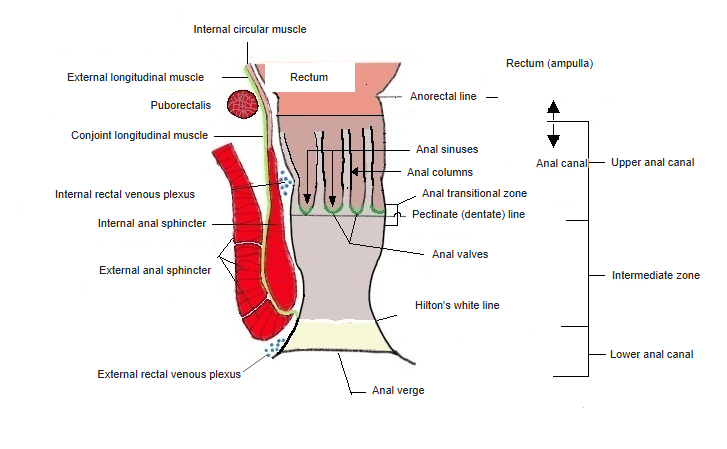
Divisions
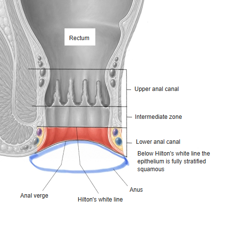
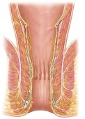
the anal canal
- Upper anal canal (= zone of the anal columns) : range: anorectal line → pectinate (dentate) line; simple columnar epithelium continuing from the rectal mucosa; length about 1.5–2 cm. Structures: anal columns, anal sinuses, anal valves.
- Intermediate zone (= transitional zone) : range: pectinate line → (Hilton's) white line; the epithelium changes to stratified squamous; length about 1–1.5 cm. The surface is covered partly by columnar and partly by stratified squamous epithelium; a venous plexus is well developed under the mucosa.
- Lower anal canal (= skin zone) : range: (Hilton's) white line → anal verge; length from a few mm to 1 cm. The surface is covered by stratified squamous epithelium rich in melanin pigment; at its end is the anus, whose edge is the anal verge; sweat glands are also found.
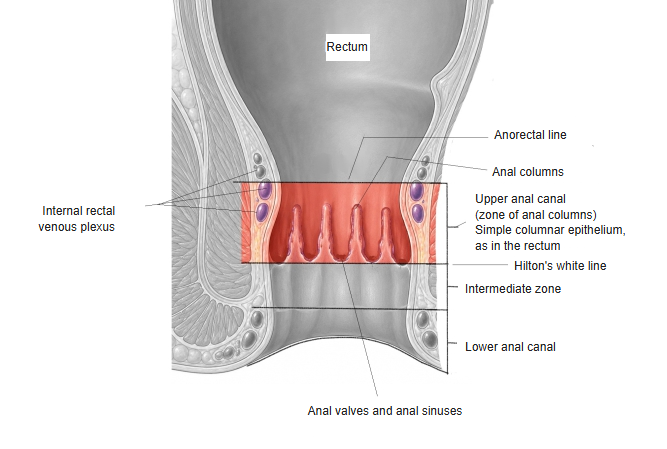
■ Upper anal canal (= zone of the anal columns) ■
· The part continuing from the rectum at the anorectal line (not a visible line); the uppermost part of the anal canal.
· Range: from the anorectal line to the pectinate line.
· Epithelium: simple columnar epithelium, as in the rectum.
· Vessels: a rich venous plexus (internal hemorrhoidal venous plexus) lies outside it.
· Structures: all of the following run round the whole lumen of the anal canal.
1. Anal columns: 6–10 longitudinal ridges.
2. Anal sinuses: pocket-like spaces at the bases of the anal columns; the anal glands open into them.
3. Anal valves: parts projecting into the lumen that form the anal sinuses. Although called "valves", they do not open and close.
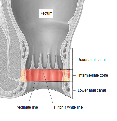
■ Intermediate zone ■
· The part continuing from the upper anal canal at the jagged pectinate line; the middle part of the anal canal.
· Range: from the pectinate line to Hilton's white line.
· Epithelium: here the simple columnar epithelium of the upper anal canal is replaced by stratified squamous epithelium, so both are present.
■ Lower anal canal ■
· The part continuing from the intermediate zone at Hilton's white line.
· Range: from Hilton's white line to the anus.
· Epithelium: completely replaced by stratified squamous epithelium, as in the skin.
· Vessels: a venous plexus (external hemorrhoidal venous plexus) lies outside it.
Structure
The differences from the wall of the rectum are summarised in the table below.
| Layer | Rectum | Anal canal | Main point of difference | |
|---|---|---|---|---|
| 1 | Mucosal epithelium | simple columnar epithelium | upper: simple columnar epithelium lower: stratified squamous epithelium | Changes to a tough structure (stratified squamous epithelium) to withstand friction as faeces pass. |
| 2 | Submucosa | relatively simple | well-developed venous plexus | The network of veins gathers and acts as a cushion. |
| 3 | Muscle layer | two layers (inner circular, outer longitudinal) | the conjoint longitudinal muscle runs between the internal and external anal sphincters | A muscle under voluntary control (external anal sphincter) is added, so defecation can be held back. |
| 4 | Serosa / adventitia | serosa: present | serosa: absent; adventitia | Lying outside the peritoneal cavity, it has no serosa; instead it is covered by adventitia, which joins it to the surrounding tissues. |
Vessels and nerves
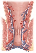
[Arteries]
The rectum and anal canal receive oxygen and nutrients mainly through the following 3 routes.
1. Superior rectal artery: the upper and middle rectum and the upper anal canal (above the pectinate line); a branch of the inferior mesenteric artery.
2. Middle rectal artery: around the lower rectum (ampulla of the rectum); a branch of the internal iliac artery.
3. Inferior rectal artery: the intermediate zone and lower anal canal (below the pectinate line); a branch of the internal pudendal artery.
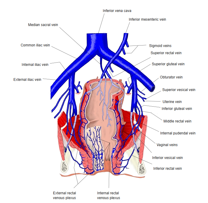
[Veins]
The veins accompany the arteries.
1. Superior rectal vein: above the pectinate line; drains through the portal vein to the liver.
2. Middle and inferior rectal veins: below the pectinate line; they do not pass through the liver but return straight to the heart through the inferior vena cava.
3. Internal / external hemorrhoidal venous plexuses
[Lymphatics]
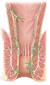
The direction of lymph flow is opposite above and below the pectinate line.
■ Route from above the pectinate line ■
Along the blood vessels (superior rectal artery), lymph flows "upwards" (towards the abdomen), deep into the abdomen: to the inferior mesenteric lymph nodes and the nodes around the aorta.
■ Route from below the pectinate line ■
Lymph flows "downwards" (towards the groin and perineum): to the inguinal lymph nodes at the groin and the internal iliac lymph nodes on the wall of the pelvis.
[Nerves]
Whether pain is felt depends on whether the part lies above or below the pectinate line.
■ Above the pectinate line (rectum to upper anal canal) ■
Nerves: autonomic nerves (sympathetic and parasympathetic; pelvic splanchnic nerves and others).
Feature: there are no nerves that feel "pain"; instead they tell the brain of the urge to defecate (rising pressure in the anal canal).
■ Below the pectinate line (intermediate zone to lower anal canal) ■
Nerves: somatic nerves (the inferior rectal nerves, branches of the pudendal nerve).
Feature: very sensitive to "pain", the same nerves as in the skin. These nerves also carry the commands that move the external anal sphincter.
Related terms
Main anatomical terms related to the anal canal, in alphabetical order.
| A | Anal columns, Anal crypts (= anal sinuses), Anal glands, Anal pecten (= intermediate zone), Anal sinus, Anal valves, Anorectal junction, Anorectal line, Anus |
| C | Columns of Morgagni, Conjoint longitudinal muscle, Corrugator cutis ani |
| I | Intermediate zone, Internal anal sphincter |
| P | Pectinate line (dentate line) |
| S | Skin zone (= lower anal canal) |
| T | Transitional zone (= intermediate zone) |
| U | Upper anal canal (= zone of the anal columns) |
References and sources
- Further reading: Anal canal, Wikipedia.