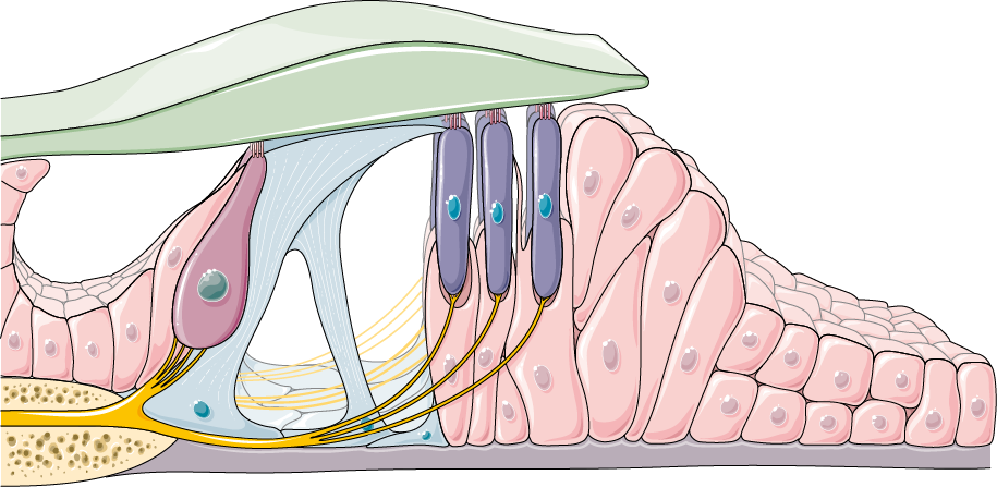
Organ of Corti
Overview
· Also called the spiral organ.
· Its underside, the lower surface of the basilar membrane, is covered by the thin cell layer that lines the scala tympani.
- Scala vestibuli : upper level of the spiral canal; filled with perilymph; passes the sound vibration on to the cochlear duct; part of the bony labyrinth.
- Vestibular membrane : (Reissner's membrane) separates the scala vestibuli from the cochlear duct.
- Cochlear duct : the middle of the three spaces, between the scala vestibuli and the scala tympani (bounded by the vestibular membrane above and the basilar membrane below); filled with endolymph and holds the spiral organ; both ends are blind. Contents: the tectorial membrane, the organ of Corti, the internal and external spiral sulci, the spiral prominence and the stria vascularis.
- Osseous spiral lamina + basilar membrane : (with the secondary spiral lamina) separate the cochlear duct and the scala vestibuli above from the scala tympani below.
- Scala tympani : lower level; part of the bony labyrinth, filled with perilymph; carries the travelling wave to the round window. It joins the scala vestibuli at the helicotrema.
- Modiolus : the cone of spongy bone at the centre of the cochlea, with the longitudinal canals and the spiral canal of the modiolus.
| 1 | Arcuate zone | · Also called the covered zone. · The part nearer the modiolus; it is tall and covered by the tectorial membrane. · The most important part of the hearing organ, made of hair cells and supporting cells. | 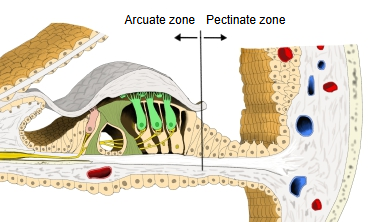 |
| 2 | Pectinate zone | · The part outside the arcuate zone. · Made of low epithelial cells (the cells of Claudius). |
Transmission of sound
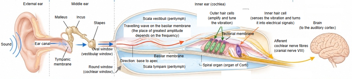
1. From the tympanic membrane to the oval window (the travelling wave starts): sound is collected by the external ear and makes the tympanic membrane vibrate. The vibration is amplified by the auditory ossicles (malleus, incus and stapes), and the movement of the stapes reaches the perilymph of the cochlea through the oval window (vestibular window). This sets up a travelling wave on the basilar membrane.
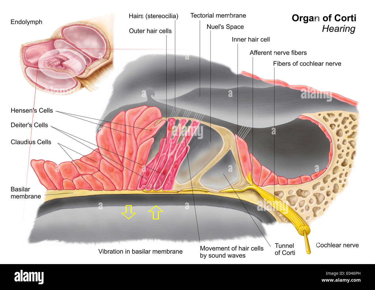
2. Vibration of the basilar membrane and stimulation of the spiral organ: the travelling wave runs from the base of the cochlea towards the apex, and the basilar membrane vibrates most at a place that depends on the frequency of the sound. This vibration makes the basilar membrane and the tectorial membrane shear against each other, so the hairs (stereocilia) of the hair cells of the spiral organ (organ of Corti) are bent: a mechanical stimulus.
3. Role of the hair cells and transmission to the nerve: the shearing bends the stereocilia of the outer and inner hair cells at the same time. The outer hair cells, which can change their length, amplify and tune the vibration of the basilar membrane (the cochlear amplifier). The inner hair cells turn the mechanical stimulus into electrical signals, which go mainly from the inner hair cells along the afferent fibres of the cochlear nerve to the brain (auditory cortex).
Points
· The sound vibration reaches the outer and inner hair cells together (in parallel), as a vibration of the basilar membrane.
· The outer hair cells mainly amplify and tune the vibration; the inner hair cells are mainly the sensory receptors.
· The round window (cochlear window) moves in the opposite phase to the oval window and so releases the pressure of the perilymph.
⇒ See also sound conduction in the cochlea.
When sound arrives, the basilar membrane vibrates first and the whole sensory epithelium moves with it. The hairs (stereocilia) of the hair cells bend, and mechanically gated ion channels at their tips open. Potassium ions, plentiful in the endolymph, flow through these channels into the hair cell and excite it electrically. In this step the mechanical stimulus of sound is turned into an electrical signal. The hair cell then releases its neurotransmitter, and the sound information travels to the brain along the nerve fibres.
 Organ of Corti
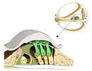 Spiral organ (section)
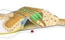 Spiral organ (3D view)
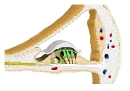 Cochlear duct (section)
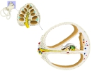 Section of a cochlear turn
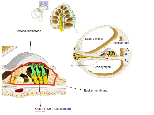
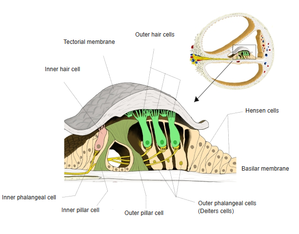
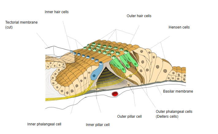
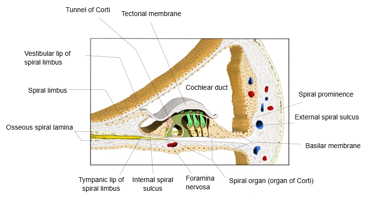
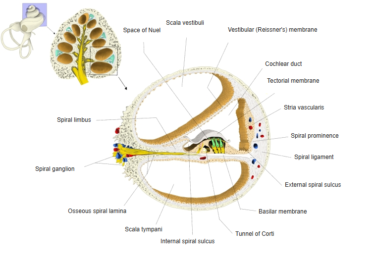
Cells of the organ of Corti
The cells that make up the organ of Corti can be divided broadly into hair cells and supporting cells.
| 1 | Hair cells | a | Inner hair cell | · Lie in a single row inside the inner pillar cells (on the modiolus side). · There are said to be about 3,500. |
| b | Outer hair cell | · Lie in several rows (three to five) outside the outer pillar cells. · Similar in shape to the inner hair cells, but nearly four times as many. · Their upper ends are held in the holes of the reticular membrane. | ||
| 2 | Supporting cells | a | Inner pillar cell | · Lies just outside the inner hair cells. · Forms the tunnel of Corti with the outer pillar cells. |
| b | Outer pillar cell | · Lies inside the outer hair cells. · Forms the tunnel of Corti with the inner pillar cells. · Bounds the space of Nuel on its inner side, with the outer hair cells and the outer phalangeal cells on its outer side. | ||
| c | Inner phalangeal cell | · Lies inside the inner pillar cells and supports the inner hair cells. | ||
| d | Outer phalangeal cell | = cell of Deiters · Cells in two or three rows under the outer hair cells. · The very thin upper half is the phalangeal process. · With the outer hair cells, they form the outer wall of the space of Nuel, whose inner wall is the outer pillar cells. | ||
| e | Cell of Hensen | · Cells in several rows outside the outer phalangeal cells, forming the sloping outer part of the organ of Corti. |
Reference websites
[Wikipedia]
· Organ of Corti, Wikipedia.
· Cochlear amplifier, Wikipedia.
[Websites with illustrations]
· Cross-section of the cochlea (figure) (Wikimedia Commons)

· Organ of Corti in cross-section (figure) (Shutterstock)

· Anatomy of the organ of Corti (figure) (Alamy)
· Organ of Corti (drawing) (Servier Medical Art)