Primary urine (Latin: urina primaria)
Also called the glomerular filtrate or ultrafiltrate
- Overview
- Route
- Composition
- Volume and changes in composition
- The three filtering layers
- Reference websites
- (Wikipedia, websites with illustrations)
- Related terms
Overview
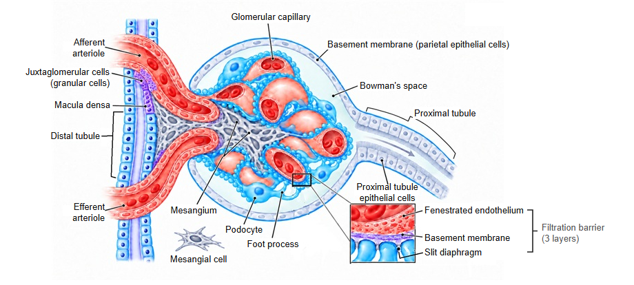
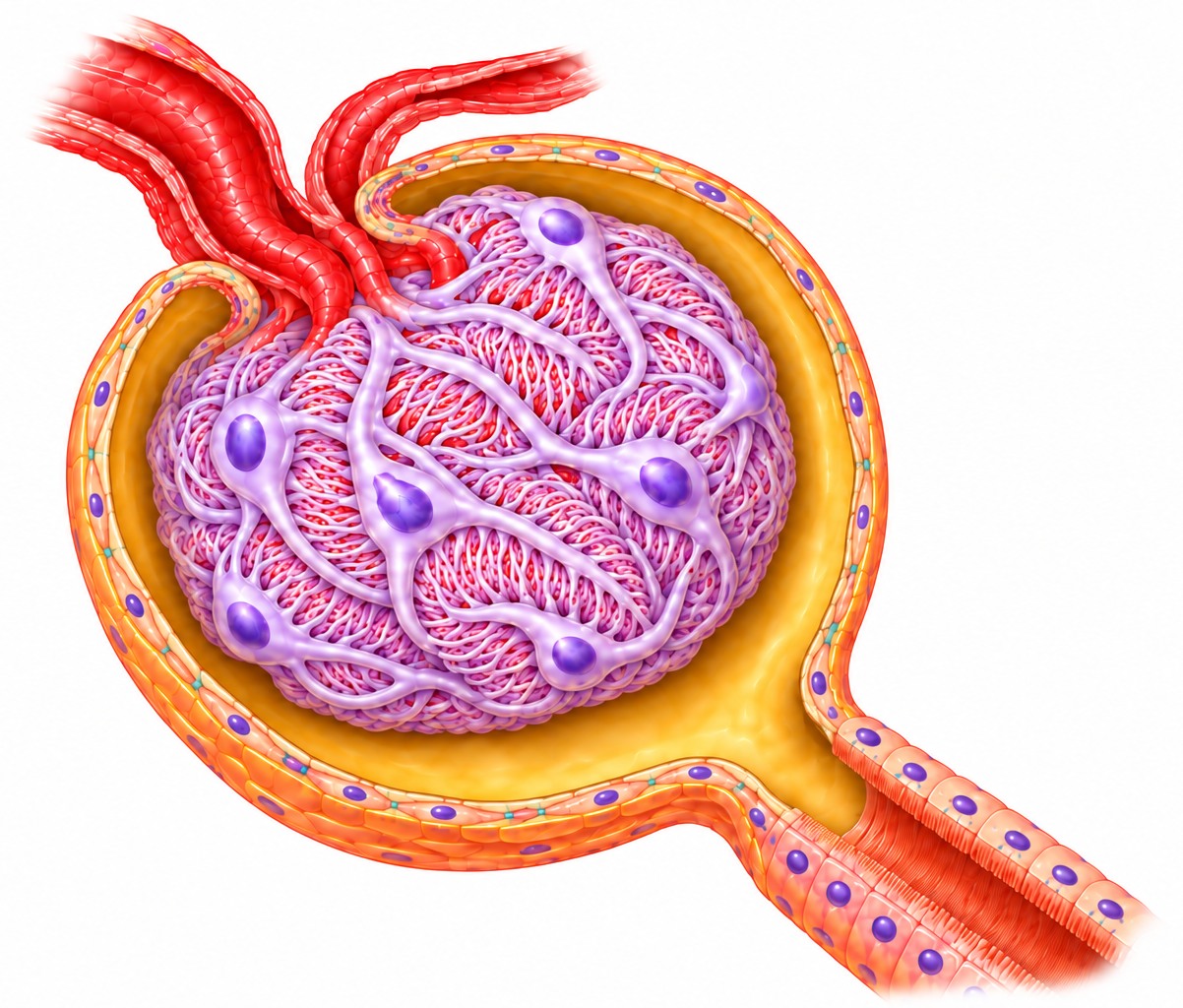
· Primary urine is the fluid filtered out of the blood in the glomerulus of the renal corpuscle. It collects in the capsular space (Bowman's space) inside the glomerular (Bowman's) capsule. It is the starting material of urine and is also called the glomerular filtrate.
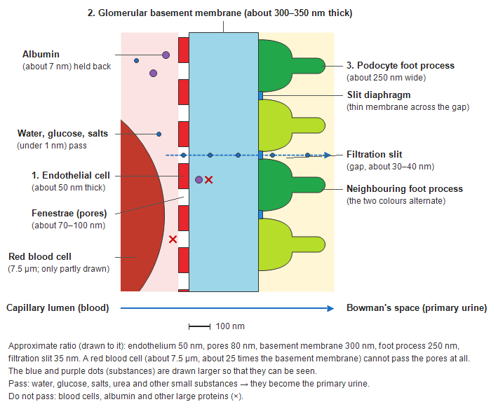
· It forms by passing three layers: the pores of the endothelial cells of the glomerular capillaries, the glomerular basement membrane, and the gaps between the processes of the podocytes (the filtration slits).
· The force that drives filtration is the blood pressure in the glomerular capillaries. It is higher than the colloid osmotic pressure of the plasma proteins plus the pressure in the capsular space, so water and small substances are pressed out of the blood.
Route
The primary urine made in the renal corpuscle leaves the kidney by the following route.
First, looking at the whole way from production to excretion:
(1) The primary urine made in the glomerulus flows through the fine tubes in the renal parenchyma and is collected in the renal sinus; from there it leaves the kidney and passes into the ureter.
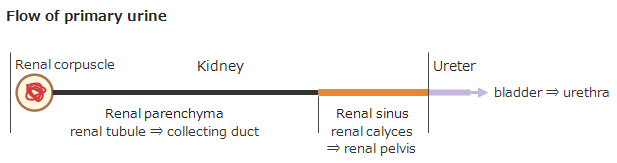
Next, looking at the flow through the renal parenchyma in more detail:
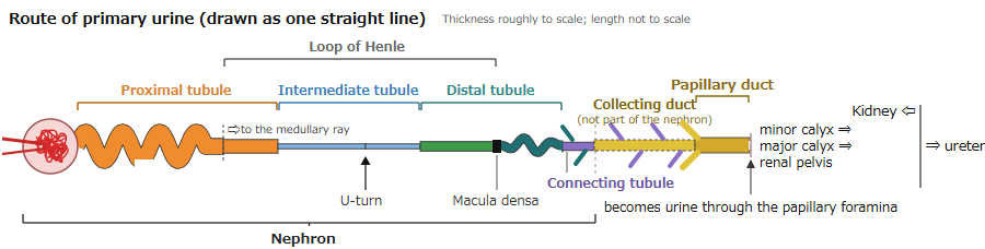
(2) The primary urine flows through the renal tubule, which alternates between coiled and straight parts, into the straight collecting duct. Just before the collecting duct lies the connecting tubule, which guides it in.
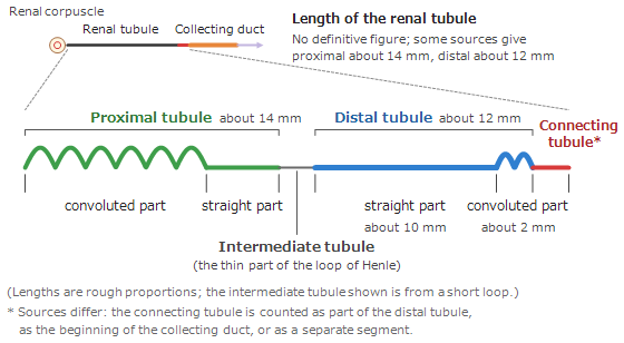
The renal tubule is drawn as a straight line above, but in reality its course from the straight part of the proximal tubule runs deep toward the renal medulla and turns back in a U-shaped loop: this is the loop of Henle (the proximal straight part, the intermediate tubule and the distal straight part; a name for the loop, not one of the divisions of the tubule).
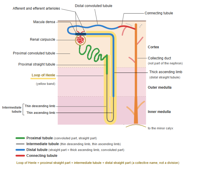
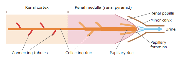
(3) From the connecting tubule the primary urine enters the collecting duct and runs straight down through the medulla toward the renal papilla. At the renal papilla, at the lower end of the collecting ducts, it leaves through the papillary foramina (the openings of the papillary ducts) into a minor calyx and becomes urine.
Summary
The way of primary urine from the renal corpuscle, where it is made, to the minor calyx, where it becomes urine, is summarized below.
Composition
· Primary urine is almost the same as blood plasma without its large proteins. Water, glucose, amino acids, electrolytes (sodium, potassium, chloride and others), urea, creatinine and uric acid are present at about the same concentrations as in plasma.
· Blood cells and plasma proteins such as albumin hardly pass through. The filter sorts molecules by their size and electric charge.
| Blood (in the glomerular capillaries) | Filter | Primary urine (Bowman's space) |
|---|---|---|
| Blood cells (red and white blood cells and others) | ✕ | do not pass |
| Proteins (albumin and others) | ✕ | hardly pass |
| Water | passes → | same as in plasma |
| Glucose, amino acids | pass → | about the same concentration as in plasma |
| Electrolytes (Na⁺, K⁺, Cl⁻ and others) | pass → | about the same concentration as in plasma |
| Waste products (urea, creatinine, uric acid) | pass → | about the same concentration as in plasma |
| Primary urine ≈ blood plasma without its large proteins | ||
| Filter = pores of the capillary endothelium → glomerular basement membrane → gaps between the podocytes (filtration slits) | ||
Volume and changes in composition
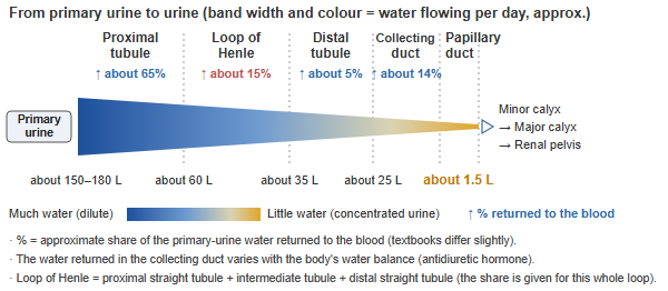
· An adult makes about 150–180 L of primary urine a day (about 100–125 mL a minute). This amount is the glomerular filtration rate (GFR), a standard measure of how well the kidneys work.
· The primary urine flows from the urinary pole into the proximal tubule and then through the intermediate tubule, the distal tubule, the connecting tubule and the collecting duct. On the way, substances the body needs are returned to the blood (reabsorption): almost all the glucose and amino acids, and about 99% of the water.
| Proximal tubule | Intermediate tubule | Distal tubule | Collecting duct | |
|---|---|---|---|---|
| Returned to the blood (reabsorption) | glucose and amino acids (almost all); Na⁺, Cl⁻, water, HCO₃⁻ | water (thin descending limb); Na⁺, Cl⁻ (thin ascending limb) | Na⁺, Cl⁻, Ca²⁺ | water (antidiuretic hormone); Na⁺ (aldosterone) |
| Added from the blood (secretion) | H⁺, drugs and others | — | — | K⁺, H⁺ |
· Glucose and amino acids are normally all taken back in the proximal tubule and do not appear in the urine.
· How much water and Na⁺ the collecting duct takes back is set by hormones according to the state of the body.
The three filtering layers
The three layers that filter the blood
Glomerular filtration
Pass: water, glucose, salts and other small substances. Do not pass: blood cells and large proteins.
Note: both kidneys together receive about 1,500–1,700 L of blood a day (about 20–25% of the blood pumped by the heart). The filtrate seeps out over the whole surface of the glomerulus; about 80% of the plasma is not filtered and leaves through the efferent arteriole.
Note: the blood leaves the glomerulus not through a vein but through the narrow efferent arteriole (a capillary bed between two arterioles, called an arterial rete mirabile; in humans found only in the glomerulus), so the pressure in the glomerulus stays about 2–3 times that of ordinary capillaries and filtration goes on.
Note: about 150–180 L of primary urine is made a day, and about 99% of it is reabsorbed in the renal tubules.
Reference websites
[Wikipedia]
· Ultrafiltration (renal), Wikipedia.
[Websites with illustrations]
· Urine formation (Visible Body)
· Your kidneys and how they work (NIDDK (NIH))
· Physiology of the nephron (Wikimedia Commons)

· Renal corpuscle (Digital Histology)
Related terms
- Urine
- Glomerulus
- Capsular space
- Glomerular capsule
- Glomerular filtration rate
- Glomerular basement membrane
- Podocyte
References and sources
- Further reading: Ultrafiltration (renal), Wikipedia.
- Federative International Programme for Anatomical Terminology (FIPAT). Terminologia Anatomica, 2nd ed. (TA2), 2019.