Proximal epiphysis of femur (Latin: epiphysis proximalis femoris)
Also called the upper end of femur
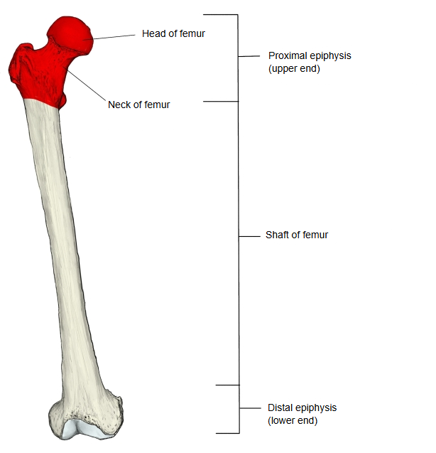
The proximal epiphysis is one of the three parts into which the femur is divided. It is less expanded than the distal epiphysis, but is marked by the neck of femur, which projects upwards and medially, and the rounded head of femur at its end. It is also called the upper end of the femur.
- Proximal epiphysis : upper end; marked by the projecting neck and head of the femur.
- Shaft of femur : the long middle part (body of femur).
- Distal epiphysis : lower end; much more expanded than the upper end.
The head of femur, smooth and covered with cartilage, projects upwards and medially; a little below and behind its centre is a small pit, the fovea of head of femur, for the ligament of head of femur. The narrower part that carries the head is the neck of femur, which meets the shaft at an obtuse angle of about 125-130 degrees in adults. Where the neck joins the shaft there are two large projections, the greater trochanter and the lesser trochanter. On the medial side of the greater trochanter is a deep pit, the trochanteric fossa, for obturator externus; obturator internus and the gemelli insert just above it. Gluteus medius, gluteus minimus and piriformis insert on the greater trochanter, and psoas major and iliacus on the lesser trochanter. In front the two trochanters are joined by the intertrochanteric line (where vastus medialis begins), and behind by the intertrochanteric crest, whose quadrate tubercle receives quadratus femoris. On the back, below the greater trochanter, is a rough area, the gluteal tuberosity, for gluteus maximus; it continues down into the lateral lip of the linea aspera. Below the lesser trochanter a ridge, the pectineal line, runs down to the linea aspera and receives pectineus.
The parts of the proximal epiphysis are listed below.
The following muscles are attached to the proximal epiphysis (red: origin; blue: insertion). Some of them continue onto the shaft of femur.
| 1 | Vastus lateralis | 2 | Vastus medialis | 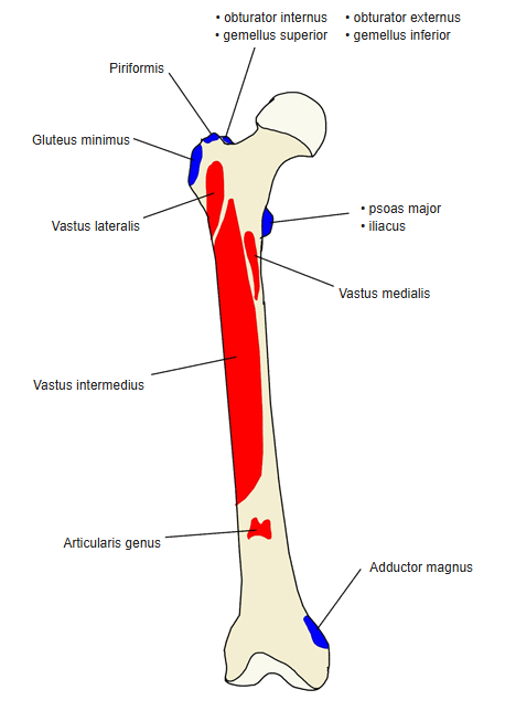 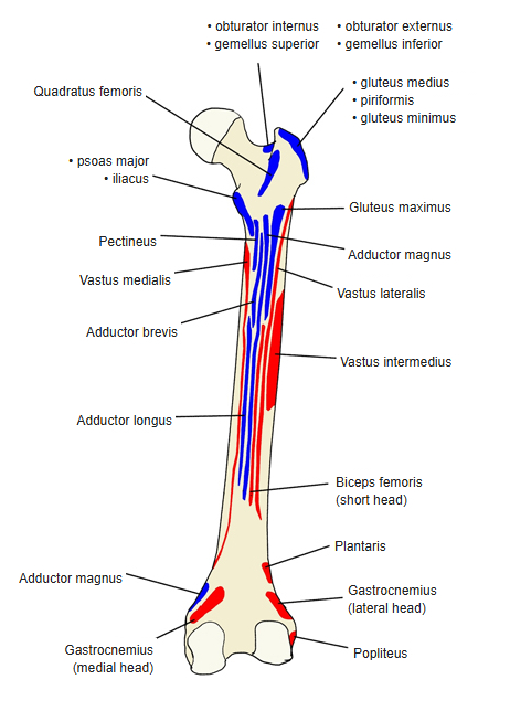 |
| 1 | Obturator externus | 6 | Psoas major | |
| 2 | Gemellus inferior | 7 | Gluteus medius | |
| 3 | Gemellus superior | 8 | Iliacus | |
| 4 | Gluteus minimus | 9 | Obturator internus | |
| 5 | Quadratus femoris | 10 | Piriformis |
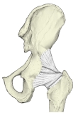
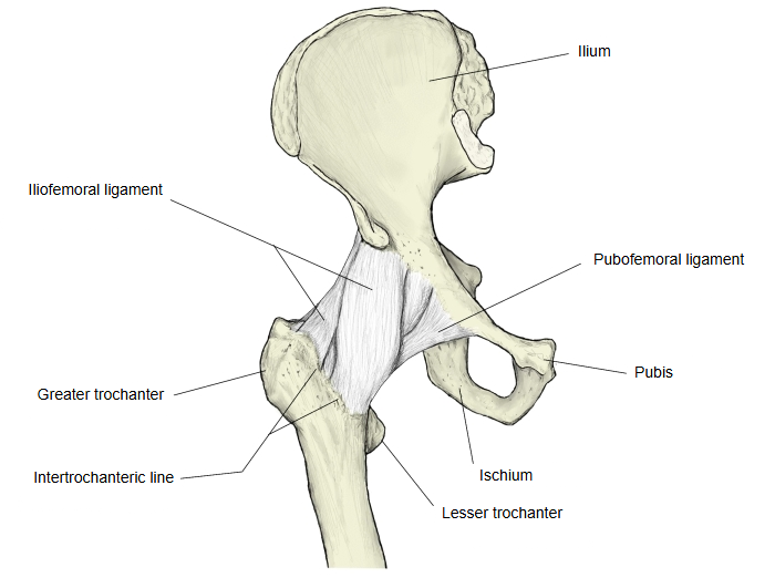
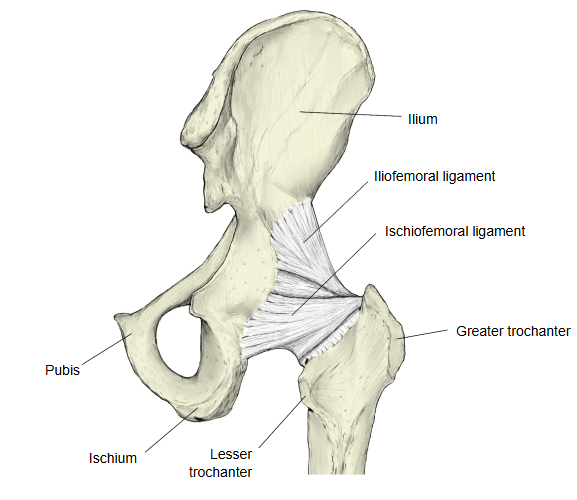
The following ligaments of the hip joint are attached to the upper femur.
| 1 | Iliofemoral ligament | Femur (greater trochanter region and intertrochanteric line) ⇔ ilium (anterior inferior iliac spine and the upper rim of the acetabulum) |
| 2 | Pubofemoral ligament | Femur (lower part of the neck, near the lesser trochanter) ⇔ pubis (iliopubic eminence and superior pubic ramus) |
| 3 | Ischiofemoral ligament | Femur (neck of femur, near the trochanteric fossa) ⇔ ischium (rim of the acetabulum) |
| 4 | Ligament of head of femur | Femur (fovea of head of femur) ⇔ hip bone (acetabular fossa) |
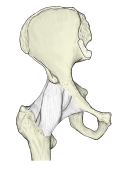 Hip ligaments (anterior)
 Hip ligaments (posterior)
Reference websites
[Wikipedia]
· Femur, Wikipedia.
· Hip, Wikipedia.
[Websites with illustrations]
· Upper end of the femur, anterior view (photograph) (Wikimedia Commons)

· Upper end of the femur, posterior view (photograph) (Wikimedia Commons)

· The femur (TeachMeAnatomy)
Where it is found
Related terms
- Head of femur
- Neck of femur
- Greater trochanter
- Lesser trochanter
- Intertrochanteric line
- Intertrochanteric crest
- Shaft of femur