Body of sphenoid (Latin: corpus ossis sphenoidalis)
· Texts count the surfaces differently: some describe four (superior, inferior, anterior and posterior), others six, adding the two lateral surfaces from which the wings arise.
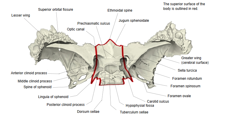
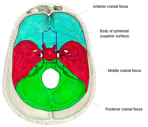
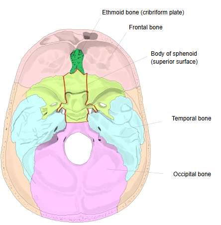
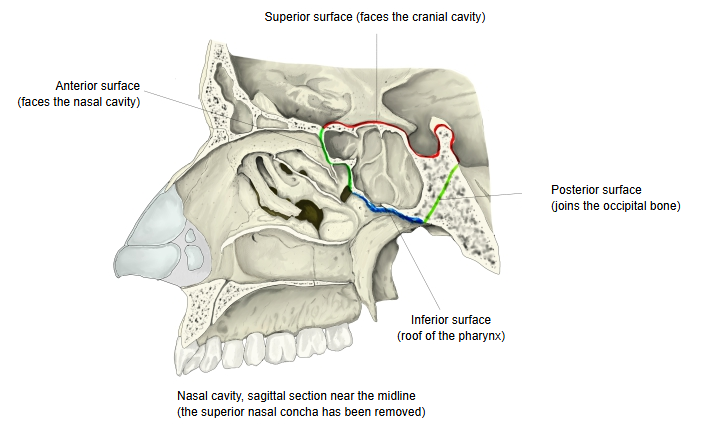
| 1 | Superior surface | Faces the cranial cavity and forms the central part of the cranial fossae. Joins: in front the ethmoid bone (cribriform plate). Behind, the back of the dorsum sellae forms the clivus together with the basilar part of the occipital bone. In the middle of the superior surface is a hollow, the sella turcica; in and around it lie the hypophysial fossa, the tuberculum sellae, the middle clinoid process, the chiasmatic groove, the dorsum sellae, the posterior clinoid process, the carotid sulcus and the sphenoidal lingula. |
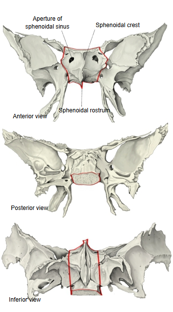
| 2 | Inferior surface | Forms the roof of the pharynx. Joins: the vomer (ala of vomer). In the midline the sphenoidal rostrum points down and forwards between the alae of the vomer. |
| 3 | Anterior surface | Faces the nasal cavity. Joins: the ethmoid bone (and the orbital processes of the palatine bones). Features: the sphenoidal crest, continuing onto the under side as the sphenoidal rostrum, and the sphenoidal concha (with the aperture of the sphenoidal sinus). |
| 4 | Posterior surface | A rough, four-sided surface. Joins: the occipital bone (basilar part). |
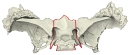 Sphenoid bone (superior view)
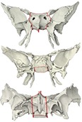 Sphenoid bone (anterior, posterior and inferior views)
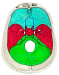 Cranial fossae 1
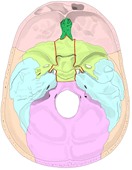 Cranial fossae 2
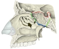 Nasal cavity (sagittal section)
· The lesser wings, the greater wings and the pterygoid processes all spring from the body.
· The septum between the two sphenoidal sinuses is often off the midline, so the two sinuses are usually unequal.
· The joint between the posterior surface and the occipital bone is a plate of cartilage in the young and turns to bone in adulthood.
The body of the sphenoid lies in the middle of the bone and is roughly cube-shaped. In the middle of its superior surface is the saddle-shaped sella turcica, with the oval hypophysial fossa in its centre. Behind the sella rises a plate of bone, the dorsum sellae, whose upper lateral corners form the posterior clinoid processes; the back of the dorsum slopes down and, with the basilar part of the occipital bone, forms the clivus. In front of the fossa a transverse ridge, the tuberculum sellae, marks the border with the front part of the body; the middle clinoid processes at its ends are small and often absent. In front of the tuberculum a narrow transverse groove, the chiasmatic groove, leads on each side to the optic canal; in front of it the smooth jugum sphenoidale is a flat surface, not a ridge. The front part of the body is joined to the lesser wings, the back part to the greater wings. Between the side of the hypophysial fossa and the root of the greater wing runs the carotid sulcus, with the plate-like sphenoidal lingula at its outer side. The inferior surface forms the roof of the nasal cavity and of the cavity of the pharynx; in its midline the sphenoidal rostrum projects down and forwards between the alae of the vomer. On the anterior surface a vertical midline ridge, the sphenoidal crest, meets the perpendicular plate of the ethmoid bone; on each side of it lies a thin sphenoidal concha, which develops as part of the ethmoid and fuses with the body during childhood, so it is most distinct in young skulls. The inside of the body is hollow, the sphenoidal sinus, divided into right and left by the septum of the sphenoidal sinuses; each half opens in front through an aperture into the sphenoethmoidal recess.
Reference websites
[Wikipedia]
· Body of sphenoid bone, Wikipedia.
· Sella turcica, Wikipedia.
[Websites with illustrations]
· Sphenoid bone: anatomy, function and development (Kenhub)
· Sphenoid bone, upper surface (Gray's Anatomy, fig. 145) (Wikimedia Commons)

· Sphenoid bone, anterior and inferior surfaces (Gray's Anatomy, fig. 146) (Wikimedia Commons)

Where it is found
Related terms
References and sources
- Further reading: Body of sphenoid bone, Wikipedia.
- Further reading: Sella turcica, Wikipedia.
- Federative International Programme for Anatomical Terminology (FIPAT). Terminologia Anatomica, 2nd ed. (TA2), 2019.