Cecum (Latin: caecum)
- Overview
- Structure (mucosa, submucosa, muscle layer, serosa)
- Functions
- Others
- Vessels and nerves (arteries, veins, lymphatics, nerves)
- Related terms
- References
Overview
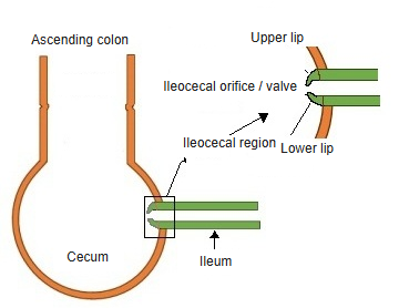
· Ileocecal region: it lies basically within the wall of the cecum (part of it projects into the lumen).
- Respiratory system
- Digestive system : oral cavity, pharynx, esophagus, stomach, small intestine, large intestine
- Urinary system
- Genital system
- Peritoneum
- Endocrine glands
large intestine
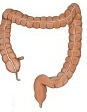
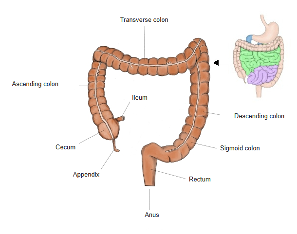
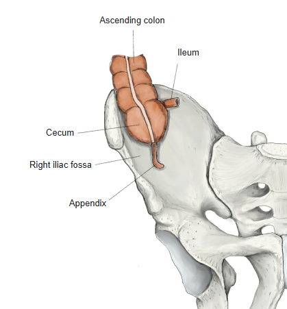
- Cecum : length 6–9 cm; the beginning of the large intestine; temporarily stores the contents from the small intestine.
- Vermiform appendix : a tube about 6–10 cm long; helps to keep the balance of the gut bacteria and takes part in immune function.
- Colon : the main part of the large intestine; functions: absorption of water and electrolytes, storage and transport of faeces. Parts: ascending colon (20–25 cm), transverse colon (40–60 cm), descending colon (20–30 cm), sigmoid colon (30–40 cm).
- Rectum : length 12–15 cm; stores faeces temporarily and passes them to the anal canal for defecation.
- Anal canal : length 3–4 cm; the last part of the digestive tract; faeces are expelled through its end, the anus.
Structure
The structure of the cecal wall is summarized below.
the cecal wall
- Mucosa (forms no circular folds)
- Epithelium : simple columnar epithelium (absorptive cells + goblet cells); intestinal crypts present, villi absent.
- Lamina propria : contains many immune cells (lymphocytes, plasma cells and others).
- Muscularis mucosae : a thin layer of smooth muscle, often with distinguishable inner circular and outer longitudinal parts.
- Submucosa : blood vessels, the submucosal plexus and lymph vessels; immunologically active in the cecum (and towards the appendix).
- Muscle layer
- Inner circular layer : smooth muscle running around the tube; thicker than the outer layer.
- Outer longitudinal layer : smooth muscle running along the tube ⇒ gathered into the three taeniae coli.
- Serosa : mesothelium (simple squamous epithelium) + thin connective tissue beneath it (blood vessels, lymph vessels, nerves and a few fat cells).
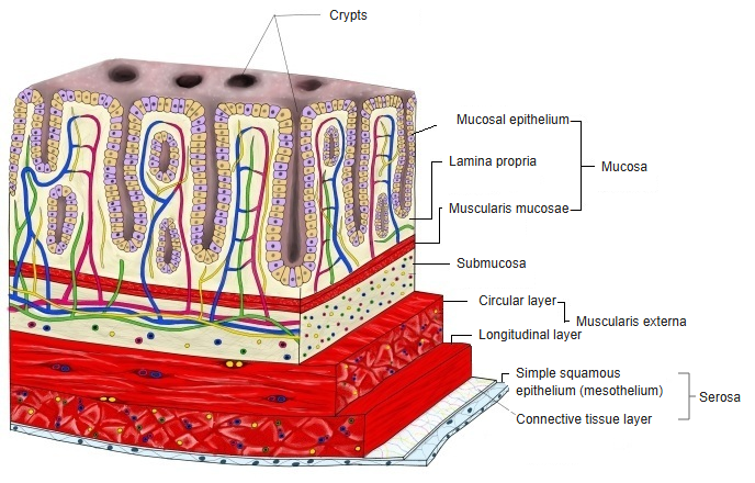
(schematic) (click to enlarge)
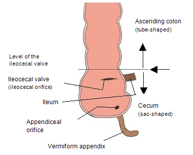
The border between the cecum and the ascending colon is indistinct, and their walls are continuous, so one cannot say exactly where one begins. One thing is certain: the ascending colon begins above the ileocecal valve. Some sources take the upper margin of the ileocecal valve as the border.
[Mucosa]
Features particular to the cecum
· Lymphoid tissue is very abundant: there are many solitary lymphoid nodules in the lamina propria.
· Its immune function is strong: it is one of the most immunologically active parts of the large intestine.
· Immunological continuity with the appendix: less than in the appendix, but gut-associated lymphoid tissue (GALT) is well developed in the cecum too.
[Submucosa]
· Lymphoid tissue is conspicuous even in the submucosa: not only in the lamina propria but also in the submucosa it is well developed.
· The density of immune cells (macrophages, lymphocytes, dendritic cells) is high.
· The character of the cecum as an "immune outpost" extends to the submucosa.
[Muscle layer]
· The muscle layer is arranged to suit a bag-shaped organ adapted to store the contents (from the ileum) for a time.
· It contributes more to mixing and holding than to peristalsis; it does not have the propulsive force of the ascending colon.
· The taeniae coli begin here: the three taeniae converge on the root of the appendix. (Taeniae coli are common to the colon, but being their starting point is peculiar to the cecum.)
[Serosa]
· As an intraperitoneal organ the cecum is highly mobile; in most cases it is covered by serosa almost all the way round.
· Its position varies greatly between individuals: high cecum, low cecum, mobile cecum.
Functions
· The cecum is the first place the contents of the small intestine enter, and, like a checkpoint, it has a well-developed immune function for defence.
Temporary storage of the contents of the small intestine (a function particular to the cecum)
Its bag-like shape suits the temporary storage of the contents coming from the small intestine. During this storage:
· gut bacteria, which are numerous from the cecum to the ascending colon, ferment and use the remaining nutrients;
· the well-developed immune system removes harmful substances.
Absorption of water and electrolytes (a function shared by the whole large intestine)
Like the rest of the large intestine, the cecum takes part in the absorption of water and electrolytes.
The distribution of gut bacteria in the large intestine is summarized below.
| Part | Amount of bacteria | Features | |
| 1 | Cecum | ◎ very many | Centre of fermentation |
| 2 | Ascending colon | ◎ many | Active fermentation |
| 3 | Transverse colon | ○ many | Stable |
| 4 | Descending colon | ○ somewhat fewer | Less water |
| 5 | Sigmoid colon, rectum | △ fewer | Preparing for defecation |
Others
In about 18% of people the cecum lies further to the left than usual, sometimes reaching or crossing the midline, and at the same time a little lower; in about 12% it is displaced upwards and to the right (mobile cecum). The cecum may lie higher than normal; in extreme cases the ascending colon is almost absent. In pregnancy the cecum, the appendix and the ascending colon are often pushed upwards by the enlarged uterus.
Vessels and nerves
[Arteries]
artery
- Ascending (colic) branch : runs upwards, giving branches to the ascending colon. Anastomosis: a branch of the right colic artery.
- Anterior cecal branch : supplies the front of the cecum.
- Posterior cecal branch : supplies the back of the cecum; often variable.
- Appendicular artery : supplies the appendix; often variable.
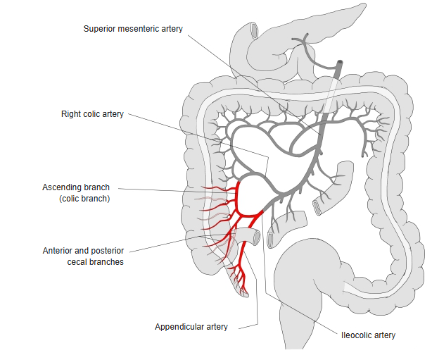
[Veins]
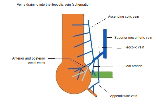
ileocolic vein (click to enlarge)
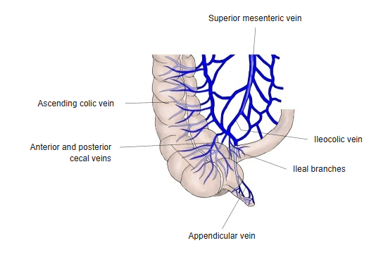
ileocolic vein
- Ileal branch : collects blood from the terminal ileum (especially near the ileocecal region).
- Ascending colic branch : collects blood from the proximal ascending colon.
- Anterior cecal vein : collects blood from the front of the cecum.
- Posterior cecal vein : collects blood from the back of the cecum.
- Appendicular vein : collects blood from the appendix.
· Features particular to the cecum: venous plexuses tend to be well developed (because of the bag-like shape and the stagnant contents), and the cecal veins join closely with the appendicular vein.
[Lymphatics]
Basic flow of lymph: within the intestinal wall ⇒ within the mesentery ⇒ central nodes.
the cecal wall
- Mucosal lymph vessels : in the lamina propria; well developed around the lymphoid nodules (lymphoid follicles).
- Submucosal lymph vessels : in the submucosa; fairly large vessels that collect lymph from the mucosal vessels.
- Intramuscular lymph vessels : in the muscle layer, between the circular and longitudinal layers; pass on to the lymph vessels outside the wall.
Lymph flow outside the cecal wall
As a rule, lymph from the cecum flows along the blood vessels.
· The central lymph nodes for the cecum are the ileocolic lymph nodes.
[Nerves]
The cecum is part of the large intestine, so its basic innervation follows that of the large intestine. Its autonomic fibres reach it through the superior mesenteric plexus along the ileocolic artery.
large intestine
- Enteric nervous system : the main nerve supply of the intestine, working fairly independently of the central nervous system.
- Myenteric plexus : between the inner circular and outer longitudinal muscle layers = Auerbach's plexus.
- Submucosal plexus : in the submucosa = Meissner's plexus.
- Autonomic nervous system : mainly synapses with the enteric nervous system; some fibres act directly on vessels and sphincters.
- Sympathetic : thoracic and lumbar splanchnic nerves; inhibit peristalsis and secretion, constrict vessels and sphincters.
- Parasympathetic : vagus nerve (proximal 2/3) and pelvic splanchnic nerves (distal 1/3); promote peristalsis and secretion and increase blood flow.
- Sensory nerves : part of the peripheral nervous system, running with the autonomic nerves to all layers of the wall (especially the muscle layer and serosa); concerned with pain.
- With the sympathetic nerves : visceral afferent fibres conveying pain, ischaemia and inflammation to the centre.
- With the parasympathetic nerves : visceral afferent fibres conveying physiological sensations and reflex input to the centre.
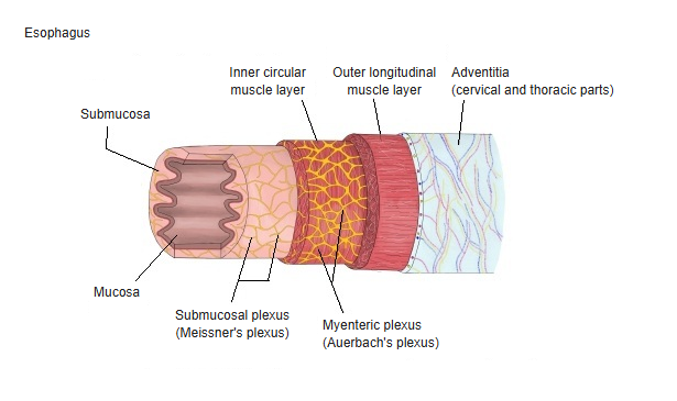
(esophagus) (click to enlarge)
Features particular to the cecum:
① The ileocecal region as a "meeting point of sensation": the junction of the terminal ileum and the cecum, where the nature of the contents changes abruptly, so visceral sensation tends to concentrate here.
② Functional unity with the nerves of the appendix: the nerves of the appendix are continuous with the cecal plexus, and the cecum itself can be the site of symptoms of appendicitis.
Related terms
Main anatomical terms related to the cecum, in alphabetical order.
| F | Frenulum of ileocecal valve |
| I | Ileocecal orifice, Ileocecal region, Ileocecal valve |
| M | Mesocecum |
| T | Taeniae coli |
| V | Vermiform appendix |
References and sources
- Further reading: Cecum, Wikipedia.