Duodenum (Latin: duodenum)
- Overview
- Position
- Parts and flexures (superior part, descending part, horizontal part, ascending part)
- Structure of the wall
- Functions
- Vessels and nerves
- Related terms
- References
Overview
· It continues from the pylorus of the stomach and forms the beginning of the small intestine.
· The name comes from the Latin duodeni, "twelve each": its length was said to equal the breadth of twelve fingers laid side by side.
- Respiratory system
- Digestive system : oral cavity, pharynx, esophagus, stomach, small intestine, large intestine
- Urinary system
- Genital system
- Peritoneum
- Endocrine glands
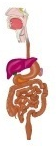
Most of the duodenum loses its mesentery during development and is fixed to the posterior wall of the abdominal cavity by the peritoneum that covers its front (a retroperitoneal organ).
· It has no mesentery, is one of the retroperitoneal organs, and receives the openings of the bile duct and the pancreatic duct; in these ways it differs from the rest of the small intestine.
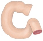 Duodenum as a whole
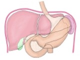 Organs below
the diaphragm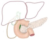 Duodenum and pancreas
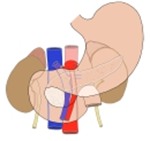 Organs and vessels
around the pancreas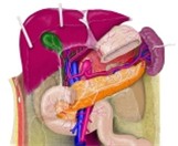 Organs around
the pancreas Trunk
(sagittal section)
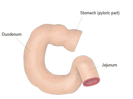
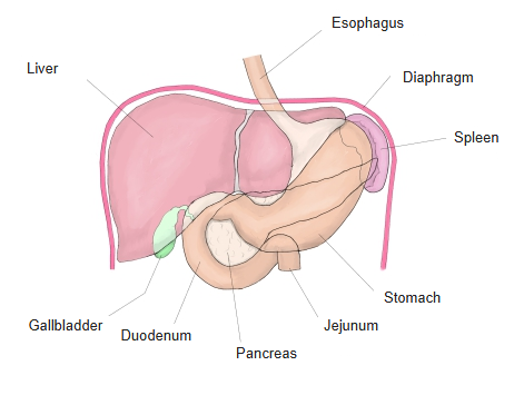
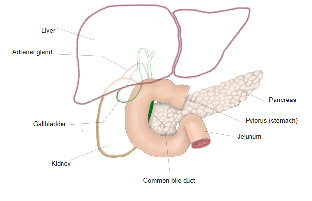
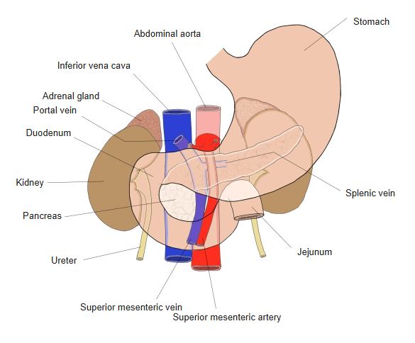
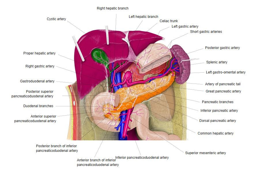
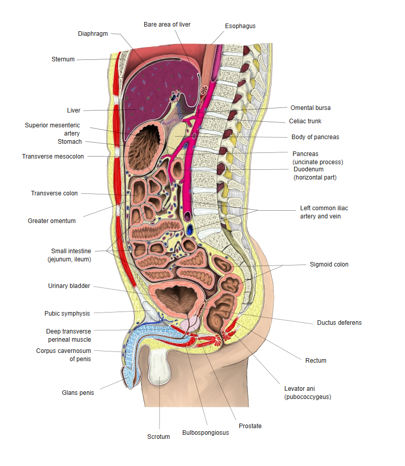
Position
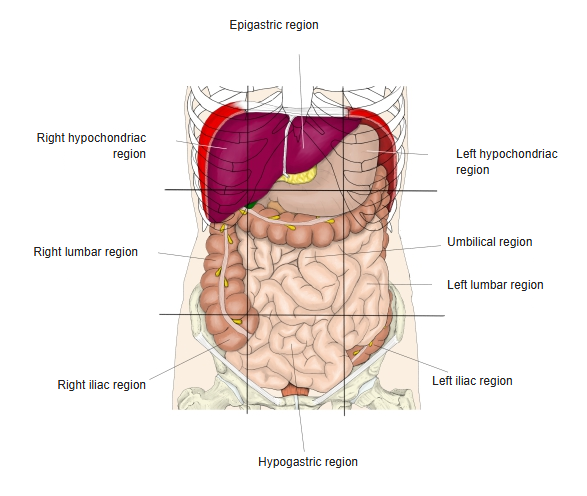
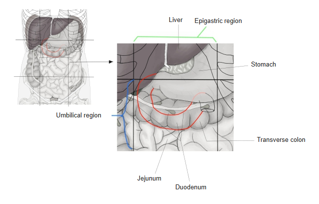
1. Position in the body
The duodenum lies in the umbilical region of the abdomen, with a part of it in the middle part of the upper abdomen.
2. Position relative to the skeleton
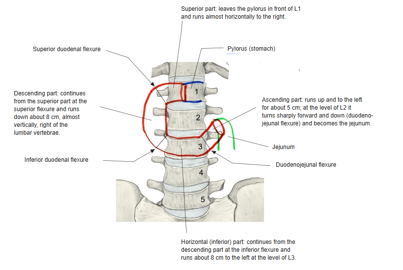
It extends from the level of the 1st to the 3rd lumbar vertebral bodies. The descending part touches the right side of the vertebral bodies, and the horizontal part runs obliquely upwards to the left of the 2nd–3rd lumbar vertebral bodies. The duodenojejunal flexure lies to the left of the 2nd lumbar vertebra.
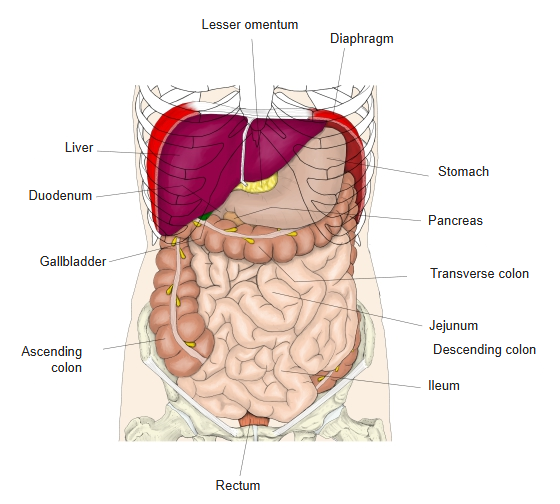
3. Relations to the surrounding organs
· It touches the liver, the gallbladder, the right kidney and the right suprarenal gland, and embraces the head of the pancreas.
· It passes in front of the inferior vena cava and the abdominal aorta and is crossed by the superior mesenteric artery and vein.
· Behind the superior part lie the portal vein, the common bile duct and the gastroduodenal artery.
· The root of the transverse mesocolon crosses the front of the descending part.
· In front of the horizontal part, the root of the mesentery encloses the superior mesenteric artery and vein.
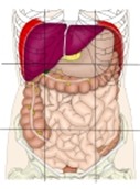 Regions of the abdomen
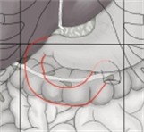 Position of the duodenum
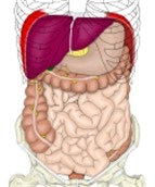 Organs of the
digestive system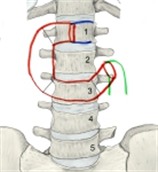 Position relative to
the skeleton
Parts and flexures
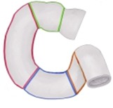
· The duodenum is divided into four parts: superior, descending, horizontal and ascending.
· It has three flexures, which form the boundaries between these parts.
flexures of the
duodenum
- Superior part : the short part (about 4–5 cm) that continues from the pylorus and runs almost horizontally.
- Superior duodenal flexure : to the right of the 1st lumbar vertebra, medial to the neck of the gallbladder.
- Descending part : descends almost vertically for about 8 cm in front of the medial border of the right kidney; bile and pancreatic juice enter here (at the major and minor duodenal papillae).
- Inferior duodenal flexure : at the level of the 3rd–4th lumbar vertebrae.
- Horizontal part : runs almost horizontally to the left for about 8 cm at the level of the 3rd lumbar vertebra.
- Ascending part : runs up and to the left for about 5 cm.
- Duodenojejunal flexure : held by the suspensory muscle of the duodenum (ligament of Treitz), connective tissue containing smooth muscle that comes from the lumbar part of the diaphragm.
 Parts and flexures
of the duodenum
[Superior part]
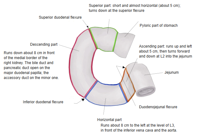
· The short part (about 4–5 cm) that continues from the pylorus of the stomach and runs almost horizontally to the right of the 1st lumbar vertebra.
· Its first 2.5 cm or so is mobile; the lesser omentum is attached to its upper border.
· Near the neck of the gallbladder it bends downwards at the superior duodenal flexure and becomes the descending part.
· Its first part is called the duodenal bulb because it looks round on radiographs; it is also the commonest site of duodenal ulcer.
[Descending part]
· Begins at the superior duodenal flexure and descends almost vertically for about 8 cm in front of the medial border of the right kidney (to the right of the vertebral column).
· At the level of the 3rd–4th lumbar vertebrae it turns sharply to the left at the inferior duodenal flexure and becomes the horizontal (inferior) part. (Some descriptions say it descends to the right of the 2nd–3rd lumbar vertebrae in front of the hilum of the right kidney.)
· Bile (through the common bile duct, opening at the major duodenal papilla) and pancreatic juice (through the pancreatic duct and the accessory pancreatic duct, opening at the major and minor duodenal papillae) are delivered here.
· Near the major duodenal papilla the lumen sometimes bulges out like a sac towards the pancreas; this is called a duodenal diverticulum.
[Horizontal part]
· At the level of the 3rd lumbar vertebra it runs almost horizontally to the left (about 8 cm) in front of the inferior vena cava and the abdominal aorta, and becomes the ascending part.
· Along the lower border of the head of the pancreas (at the level of the 3rd lumbar vertebra) it runs horizontally in front of the right psoas major, the inferior vena cava and the left psoas major.
· The superior mesenteric artery descends across the front of the horizontal part.
[Ascending part]
· Runs obliquely up and to the left (about 5 cm) and, to the left of the 2nd lumbar vertebra, turns sharply forwards and downwards (the duodenojejunal flexure) to continue as the jejunum.
· The duodenojejunal flexure is held in place by the suspensory muscle of the duodenum (ligament of Treitz), connective tissue containing smooth muscle that comes from the lumbar part of the diaphragm.
Structure of the wall
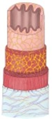
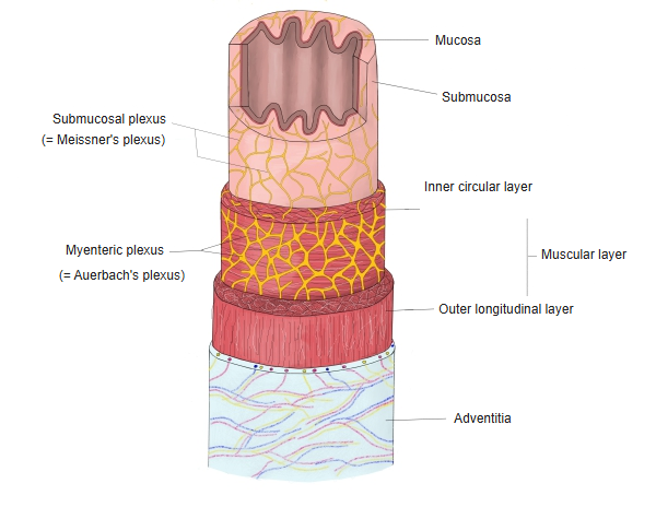
The wall of the duodenum is built on the same plan as the rest of the digestive tract.
duodenal wall
- Mucosa
- Epithelium : simple columnar epithelium for absorption and secretion; it has villi.
- Lamina propria : loose connective tissue of reticular character, with blood vessels, lymphatics and immune cells.
- Muscularis mucosae : a thin layer of smooth muscle.
- Submucosa : connective tissue containing blood vessels, nerves and the duodenal glands (Brunner's glands).
- Muscle layer
- Inner circular layer : smooth muscle.
- Outer longitudinal layer : smooth muscle.
- Outermost layer : differs by part: the superior part has a serosa, the descending and horizontal parts an adventitia, and the ascending part is partly covered by serosa.
Functions
Neutralizing the food
The contents coming from the stomach (chyme), made strongly acid by gastric acid, are neutralized by the addition of alkaline bile and pancreatic juice so that they are easier to digest.
Breaking down nutrients
· The pancreas releases pancreatic juice containing enzymes that break down proteins, fats and carbohydrates (amylase, trypsin, lipase and others).
· The liver and gallbladder deliver bile, which emulsifies fat and helps its breakdown.
Vessels and nerves
[Arteries]
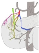
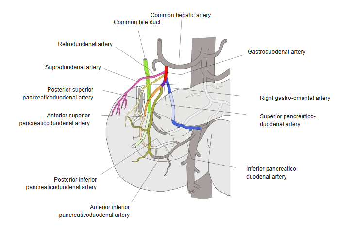
The duodenum is supplied by the following branches of the gastroduodenal artery and by the anterior and posterior inferior pancreaticoduodenal arteries, branches of the inferior pancreaticoduodenal artery.
artery
- Superior pancreaticoduodenal artery : runs between the duodenum and the head of the pancreas and divides into anterior and posterior branches. Anastomosis: inferior pancreaticoduodenal artery (anterior and posterior branches).
- Right gastro-omental artery : runs to the left along the greater curvature. Anastomosis: left gastro-omental artery. Branches: gastric and omental branches.
- Retroduodenal arteries : supply the back of the duodenum and the head of the pancreas, and give branches to the common bile duct.
- Supraduodenal artery : supplies the upper duodenum (superior and descending parts) and the head of the pancreas; it often arises from the gastroduodenal artery, but may come from the common or proper hepatic artery.
 Branches of the
gastroduodenal artery
[Veins]
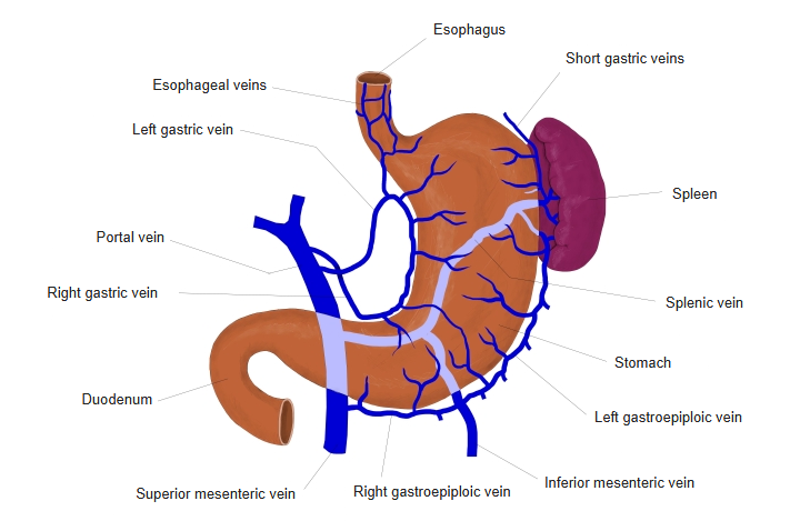
The pancreaticoduodenal veins drain into the portal vein (directly or through the superior mesenteric vein) and so return to the liver.
 Veins of the stomach
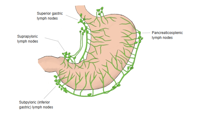
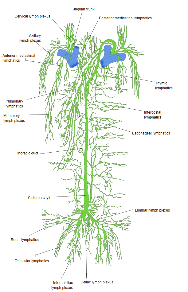
[Lymphatics]
Pancreaticoduodenal lymph nodes and superior mesenteric lymph nodes; the lymph flows on to the nodes at the porta hepatis and the superior mesenteric nodes.
 Lymph nodes around
the stomach Lymph nodes from the thoracic
to the pelvic cavity
[Nerves]
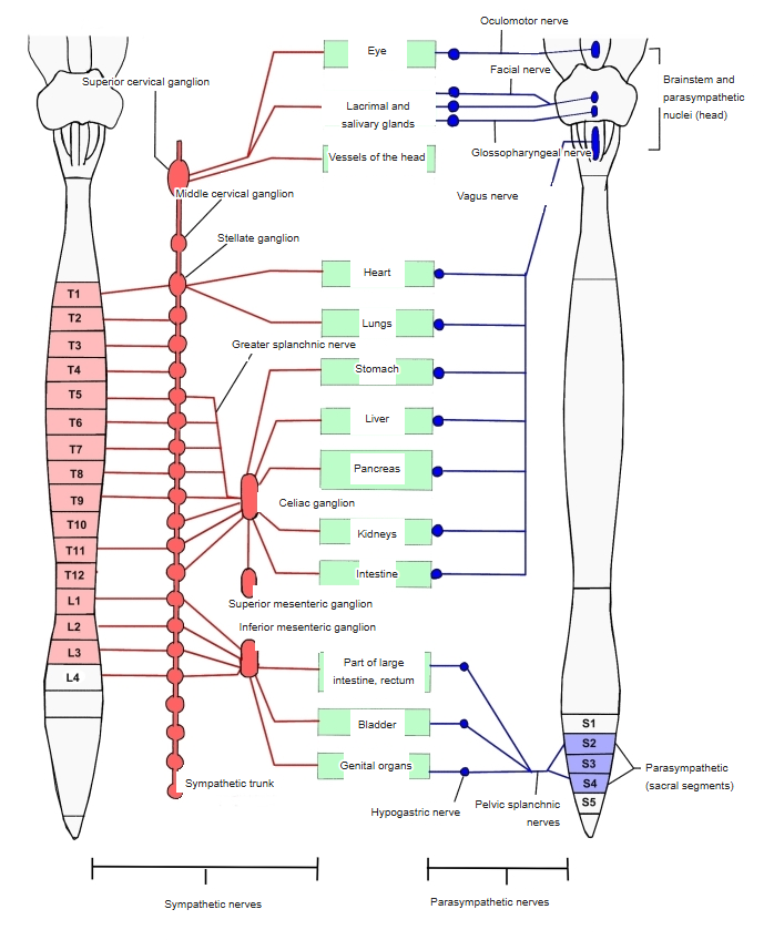
The duodenum is supplied by the autonomic nervous system.
Sympathetic nervous system
· Ganglia: celiac ganglion, superior mesenteric ganglion.
· Role: constricts the vessels (less blood flow) and inhibits peristalsis (less digestive activity).
Parasympathetic nervous system
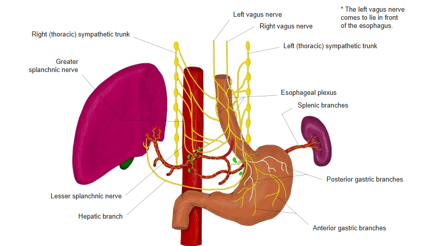
· The vagus nerve is the main parasympathetic nerve.
· Role: promotes secretion of gastric and pancreatic juice and peristalsis (more digestive activity).
Intrinsic (enteric) nervous system
· Submucosal plexus (Meissner's plexus): in the submucosa; regulates secretion of digestive juices.
· Myenteric plexus (Auerbach's plexus): between the muscle layers; controls peristalsis.
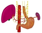 Vagus nerve
(thorax and abdomen) Autonomic nervous system
 Submucosal and
myenteric plexuses
Related terms
Main anatomical terms related to the duodenum, in alphabetical order.
References and sources
- Further reading: Duodenum, Wikipedia.