Latin: cavitas nasi
Overview
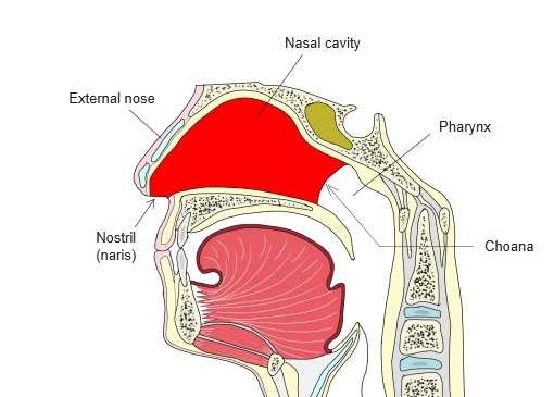
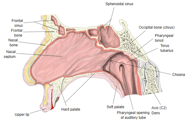
- The air space inside the nose, from the nostrils (nares) in front to the choanae behind, where it opens into the nasopharynx.
- Divided into right and left halves by the nasal septum.
- Each half has a skin-lined nasal vestibule just inside the nostril and the nasal cavity proper behind it.
- Its lining mucosa is thick and richly supplied with blood, so the airway is fairly narrow.
- Warms, moistens and filters the inhaled air, and contains the organ of smell.
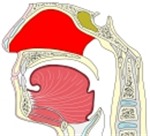
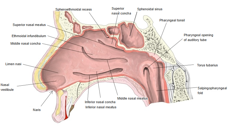
Nasal cavity (median section) 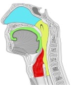
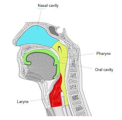
From the nasal cavity to the larynx
Bones and cartilages
| Part of the wall | Bones and cartilages |
|---|---|
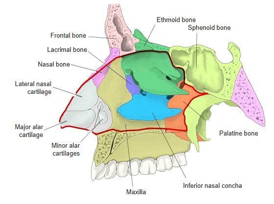
| Roof | Nasal bone, frontal bone, cribriform plate of the ethmoid, body of the sphenoid |
| Floor | Palatine process of the maxilla and horizontal plate of the palatine bone (hard palate) |
| Lateral wall | Maxilla, lacrimal bone, ethmoid (superior and middle conchae), inferior nasal concha, perpendicular plate of the palatine bone, medial pterygoid plate |
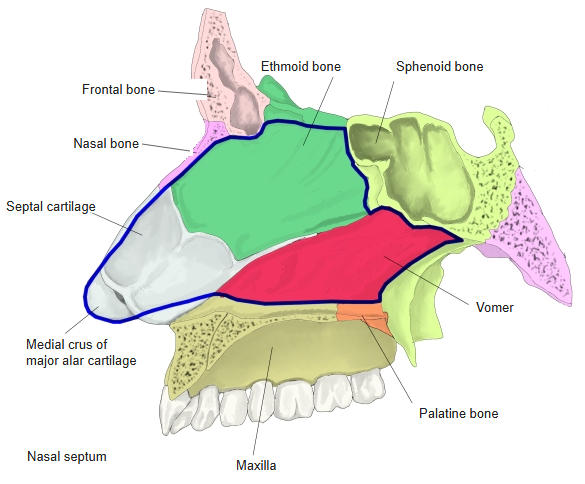
| Medial wall (nasal septum) | Perpendicular plate of the ethmoid, vomer and the septal cartilage |
| External nose (cartilages) | Lateral nasal cartilage, major alar cartilage, minor alar cartilages, septal cartilage |
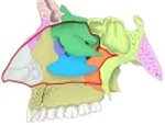
Lateral wall: bones and cartilages 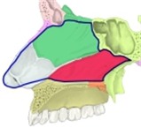
Nasal septum: bones and cartilage
Lateral wall
- The medial wall (septum) is smooth; the lateral wall is irregular.
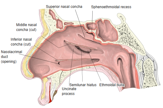
- Nasal conchae (superior, middle, inferior): three curved shelves running front to back; each overhangs a passage, the nasal meatus of the same name.
- Common nasal meatus: the narrow vertical space between the free edges of the conchae and the septum; it leads back to the choana.
- Sphenoethmoidal recess: a small space above and behind the superior concha, in front of the body of the sphenoid; the sphenoidal sinus opens here.
- Ethmoidal bulla: a rounded bulge in the middle meatus, made by the middle ethmoidal cells.
- Uncinate process: a thin curved plate of the ethmoid in front of the bulla.
- Semilunar hiatus: the curved slit between the bulla and the uncinate process; it leads forward and up into the ethmoidal infundibulum.
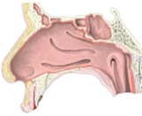
Lateral wall of the nasal cavity 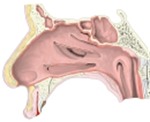
Lateral wall with the conchae removed 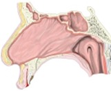
Nasal septum and nasopharynx
Nasal mucosa
- Respiratory region: most of the cavity; pseudostratified ciliated columnar epithelium with goblet cells and many nasal glands; a thick venous plexus (cavernous plexus of the conchae) warms the air.
- Olfactory region: a small area on the roof, the upper septum and the superior concha; contains the olfactory receptor cells.
- Kiesselbach area: the front of the septum, where several arteries anastomose – the usual site of nosebleeds.
Openings
| Site | What opens there |
|---|---|
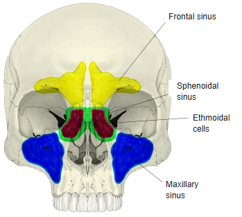
| Sphenoethmoidal recess | Sphenoidal sinus |
| Superior nasal meatus | Posterior ethmoidal cells |
| Middle nasal meatus | Frontal sinus (through the ethmoidal infundibulum), maxillary sinus, anterior and middle ethmoidal cells |
| Inferior nasal meatus | Nasolacrimal duct – about 30–35 mm behind the nostril |
| Choanae | Nasopharynx |
Paranasal sinuses
Blood supply, lymph and nerves
| Artery | Origin |
|---|---|
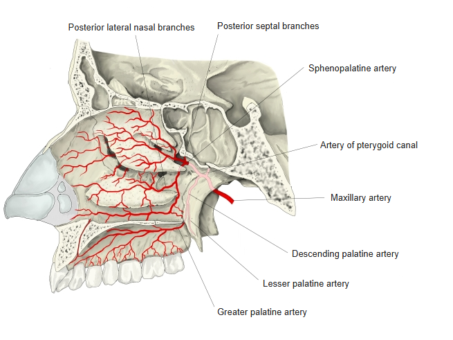
| Sphenopalatine artery (posterior lateral nasal and posterior septal branches) | Maxillary artery |
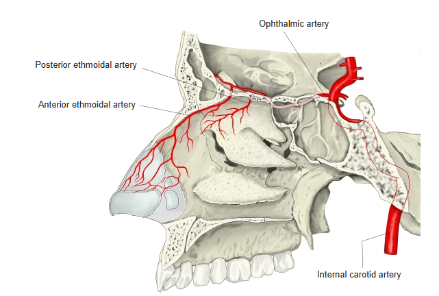
| Anterior ethmoidal artery | Ophthalmic artery (from the internal carotid artery) |
| Posterior ethmoidal artery | Ophthalmic artery |
| Greater palatine artery (through the incisive canal) | Descending palatine artery |
| Septal and alar branches | Facial artery (superior labial artery); angular artery |
- Veins: accompany the arteries and connect with veins of the face, orbit and oral cavity.
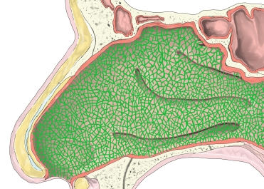
- Lymph: a network in the lamina propria drains to the retropharyngeal, deep cervical and submandibular nodes, and partly to nodes near the parotid gland.
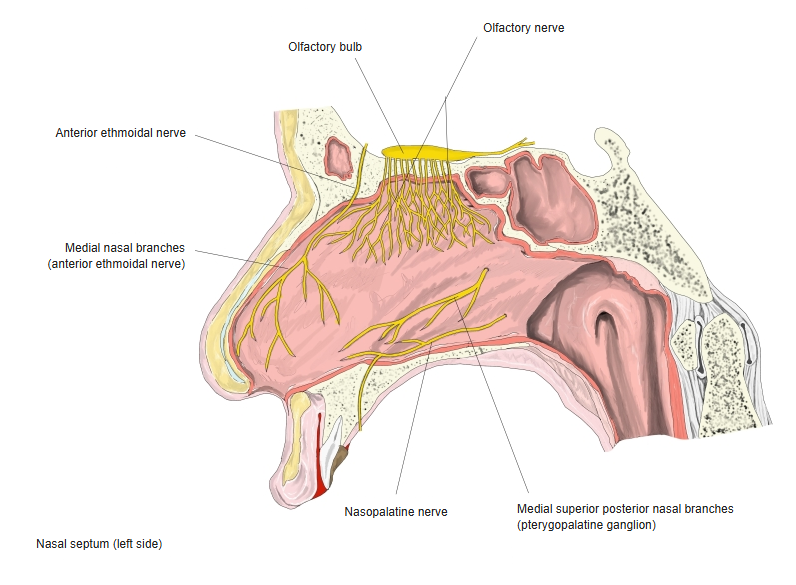
- Smell: olfactory nerves (CN I) from the olfactory region pass through the cribriform plate.
- General sensation: anterior ethmoidal nerve (from the nasociliary nerve, ophthalmic nerve) to the front; nasal branches of the pterygopalatine ganglion (maxillary nerve) to the back, including the nasopalatine nerve on the septum.
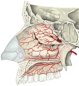
Arteries of the lateral wall (1) 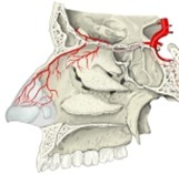
Arteries of the lateral wall (2) 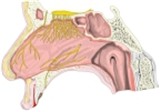
Nerves of the nasal septum 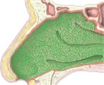
Lymph vessels of the lateral wall
References and sources
- Further reading: Nasal cavity, Wikipedia.