Teeth (Latin: dentes; singular dens)
- Overview
- Names of the parts
- Structure
- Hard tissues: dentin, enamel, cementum (acellular / cellular); dental pulp
- Two sets of teeth
- Classification and dental formula (permanent teeth, deciduous teeth, FDI system)
- Shape of the permanent teeth (surfaces and edges; incisors, canines, premolars, molars, third molars)
- Vessels and nerves
- Development
- Related terms
- References
Overview
system
- Digestive tract : takes in, digests and absorbs food and expels the residue; oral cavity → pharynx → esophagus → stomach → small intestine → large intestine
- Accessory glands : salivary glands (secreting digestive juice), digestive glands (solid organs such as the liver and pancreas)
- Others : teeth, tongue
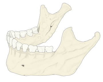
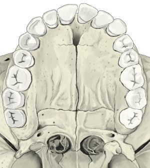
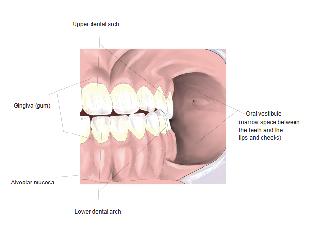
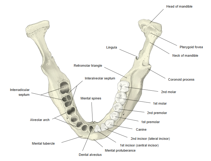
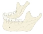 Lower dental arch
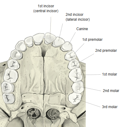
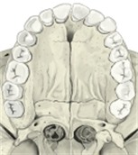 Upper dental arch
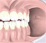 Upper and lower dental arches, gingiva
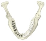 Mandible (from above)
Names of the parts
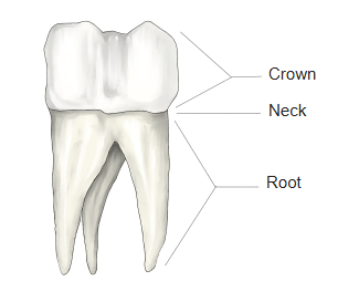
a tooth
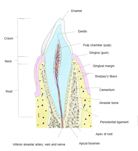
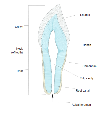
- Crown : the part that appears from the gingiva onto the surface, covered by enamel.
- Occlusal surface : the contact surface between upper and lower teeth; in the molars it is divided into cusps.
- Vestibular surface : the surface facing the oral vestibule; in the molars it is called the buccal surface.
- Lingual surface : the surface facing the oral cavity proper.
- Contact surface : the surface touching the neighbouring tooth of the same row (mesial and distal surfaces).
- Root : covered by cementum and set in the alveolus; its tip is the apex of the root.
- Neck : the slightly narrowed part at the border between crown and root.
- Pulp cavity : the inner space holding the dental pulp (rich in vessels, nerves and lymph vessels); root canal, apical foramen.
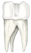 The three parts of a tooth
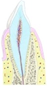 Lower incisor (sagittal section)
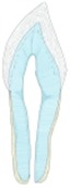 Structure of a tooth
Structure
of a tooth
- Hard tissues
- Dentin : the main material of the tooth (70%); hardness: enamel > dentin > bone.
- Enamel : the glossy substance covering the crown, the hardest material in the body; main component: hydroxyapatite (90%).
- Cementum : a bone-like layer covering the root, thicker towards the apex.
- Dental pulp
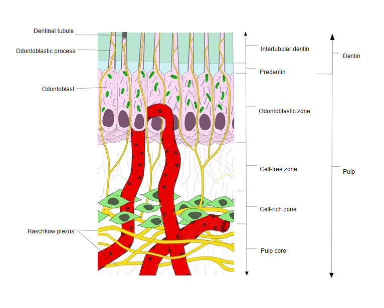
- Vessels / nerves : rich in arteries, veins, lymph vessels and nerves (forming the plexus of Raschkow).
- Cells : odontoblasts lie in the outermost layer; others: fibroblasts, undifferentiated mesenchymal cells and so on.
- Connective tissue : mainly collagen fibres(?); no elastic fibres are seen.
[Hard tissues]
tissues
- Dentin : the main component of the tooth (70%); hardness: enamel > dentin > bone.
- Enamel : the glossy substance covering the crown, the hardest material in the body; main component: hydroxyapatite (90%).
- Cementum : the bone-like layer covering the root, thicker towards the apex; acellular and cellular cementum.
- Lower incisor (sagittal section)
- Structure of a tooth
■ Dentin ■
· It contains about 70% inorganic matter and is the hardest tissue after enamel; dentin is harder than bone.
Note: the values differ between sources; some give the organic part as 30%.
· Its Mohs hardness is about 5–6, similar to glass.
The matrix of the dentin is classified as follows.
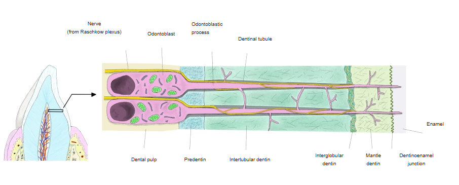
- Odontoblasts : form the dentin; the cell bodies lie in the pulp and send long processes into the dentin.
- Predentin : the innermost layer, an organic-rich zone not yet calcified.
- Dentinal fibres : = Tomes' fibres; the processes of the odontoblasts running in the dentinal tubules.
- Interglobular spaces (Owen) : irregular, poorly calcified lines running almost parallel to the outer edge of the dentin.
The following names are also used:
· Intertubular dentin: the dentin between the dentinal tubules.
· Interglobular dentin: poorly mineralised areas in the dentin, left between the calcified globules as mineralisation proceeds.
· Mantle dentin: the outermost dentin (nearest the enamel), weakly calcified.
· Dentinoenamel junction: the border between dentin and enamel.
Dentin made before the root is complete is called primary dentin; dentin made after root completion is secondary dentin. Dentin made in response to stimuli such as caries is called tertiary dentin or reparative dentin, and the secondary dentin made under normal conditions is sometimes called physiological secondary dentin to distinguish the two.
time of
formation
- Primary dentin : dentin made until the root is complete.
- Secondary dentin : dentin made after the root is complete, under normal conditions.
- Physiological secondary dentin : secondary dentin made under normal conditions.
- Tertiary / reparative dentin : dentin made in response to stimuli such as caries.
- Lower incisor (sagittal section)
- Structure of a tooth
 Structure of dentin
■ Enamel ■
tissues
- Dentin : the main component of the tooth (70%).
- Enamel : the glossy substance covering the crown, the hardest material in the body.
- Cementum : the bone-like layer covering the root.
of enamel
- Enamel rods : the main element of enamel, bundles of hydroxyapatite crystals; they run densely without branching from the dentin border towards the surface of the crown.
- Enamel sheath : the organic membrane surrounding the enamel rods.
- Interrod substance : hydroxyapatite crystals filling the narrow gaps between the rods.
- Lower incisor (sagittal section)
- Structure of a tooth
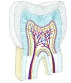 Molar (section)
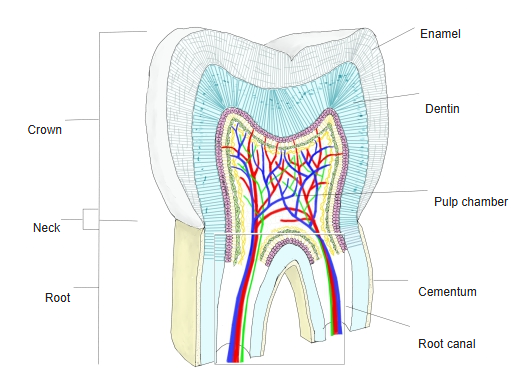
The features of enamel are briefly as follows.
· The enamel rods do not branch and run with a slight wave.
· The enamel rods and the interrod substance are strongly calcified.
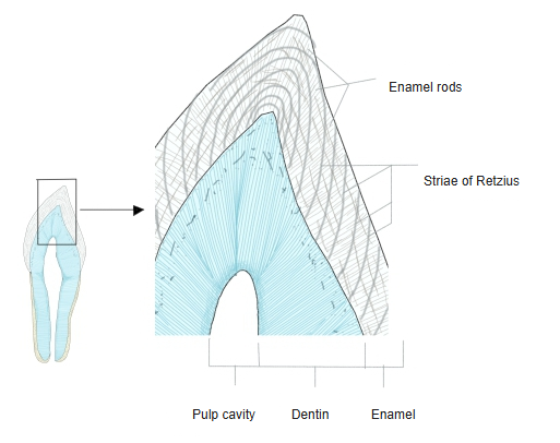
· The enamel rods have poorly calcified places at regular intervals; in sections these are seen as the striae of Retzius.
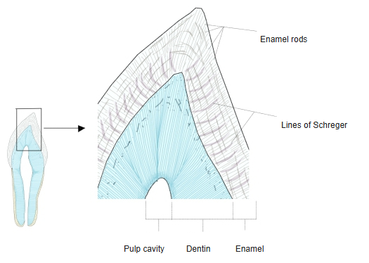
· The rods run in the same direction within each region, and the borders between neighbouring regions can be seen as the lines of Schreger.
· Just after eruption, the enamel surface has a thin, tough membrane (enamel cuticle / Nasmyth's membrane).
⟨Course of the enamel rods⟩
In ground longitudinal sections the course of the rods is not simple: places where groups of rods curve together alternate at regular intervals with places where they run almost straight. Groups of rods cut across appear as dark bands, and groups cut lengthwise as light bands, together forming alternating light and dark stripes: the lines of Schreger.
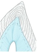 Striae of Retzius
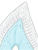 Lines of Schreger
■ Cementum ■
- Lower incisor (sagittal section)
- Structure of a tooth
- Molar (section)
Note: the figures differ between sources; take them only as a guide.
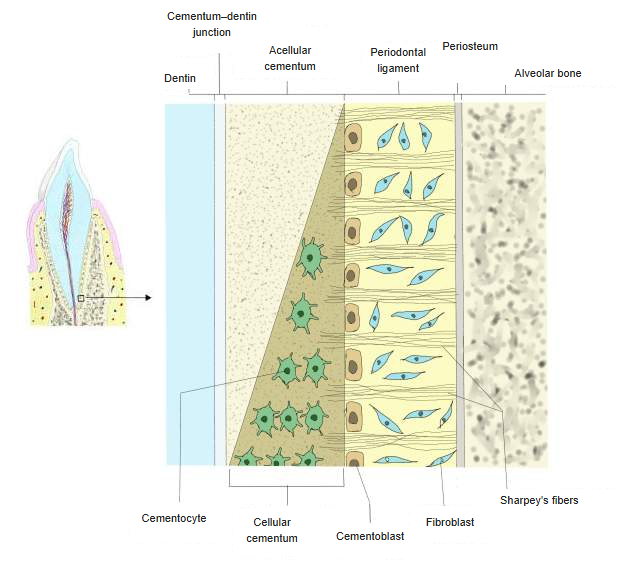
Acellular cementum
· Position: mainly covers the dentin thinly from the neck to near the apex.
· Feature: called "acellular" because it contains no cementocytes.
· Role: anchors the root firmly to the alveolar bone.
Cellular cementum
· Position: mainly in the apical third of the root, covering the acellular cementum.
· Feature: called "cellular" because it contains cementocytes.
· Role: takes part in repair and regeneration of the root; important when the root needs to grow or be repaired.
 Structure of cementum
[Dental pulp]
· Developmentally it is the grown-up dental papilla of mesodermal origin.
· It nourishes the tooth (and, in childhood, also controls its growth).
The myelinated nerves seen in the pulp come from the trigeminal nerve; they form a plexus in the pulp, from which only axons pass a little way into the dentin. A capillary network is also well developed.
- Lower incisor (sagittal section)
- Pulp cavity
- Structure of dentin
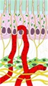 Outer layer of the dental pulp
Two sets of teeth
[Order and timing of eruption]
| Deciduous teeth (milk teeth) | Permanent teeth | ||||
|---|---|---|---|---|---|
| 1 | 1st incisor | 6–8 months | 1 | 1st molar (1) | 6–7 years |
| 2 | 2nd incisor | 7–12 months | 2 | 1st incisor | 7–8 years |
| 3 | 1st deciduous molar | 12–16 months | 3 | 2nd incisor | 8–9 years |
| 4 | Canine | 15–20 months | 4 | 1st premolar | 9–11 years |
| 5 | 2nd deciduous molar | 20–30 months | 5 | Canine | 11–13 years |
| 6 | 2nd premolar | 11–15 years | |||
| 7 | 2nd molar | 13–16 years | |||
| 8 | 3rd molar (2) | 17–40 years | |||
(1) also called the "six-year molar"; (2) also called the wisdom tooth.
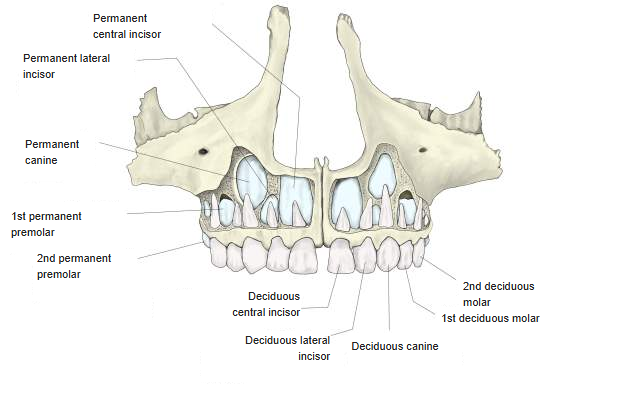
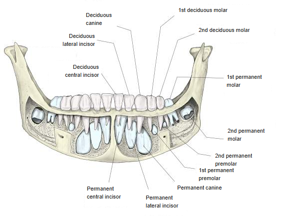
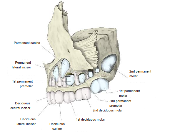
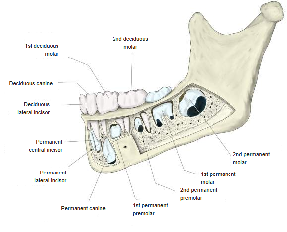
The illustrations below show the teeth of a 6-year-old child; blue shows the germs of the permanent teeth. By 6 years all the deciduous teeth have erupted and are still present, and the first permanent molar, the "six-year molar", starts to erupt at this age.
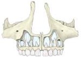 Teeth at 6 years
(upper jaw, front)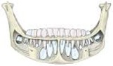 Teeth at 6 years
(lower jaw, front)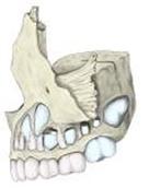 Teeth at 6 years
(upper jaw, left side)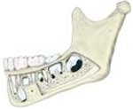 Teeth at 6 years
(lower jaw, left side)
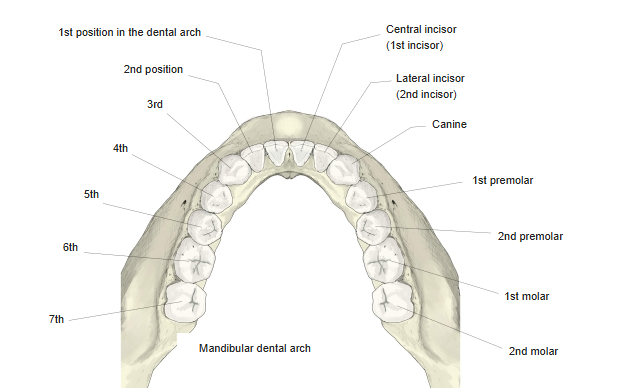
Classification and dental formula
[Classification and dental formula of the permanent teeth]
■ Classification (the same in upper and lower jaws) ■
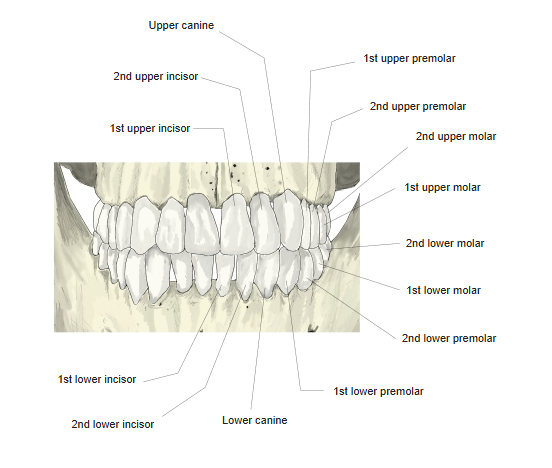
| Anterior teeth | Incisors | 1st incisor [1] |
|---|---|---|
| 2nd incisor [2] | ||
| Canine | canine [3] | |
| Posterior teeth (cheek teeth) | Premolars | 1st premolar [4] |
| 2nd premolar [5] | ||
| Molars | 1st molar [6] | |
| 2nd molar [7] | ||
| 3rd molar [8] |
■ Dental formula ■
| Used in dentistry | ||||||||||||||||
|---|---|---|---|---|---|---|---|---|---|---|---|---|---|---|---|---|
| Half (right) | Half (left) | |||||||||||||||
| Upper | 8 | 7 | 6 | 5 | 4 | 3 | 2 | 1 | 1 | 2 | 3 | 4 | 5 | 6 | 7 | 8 |
| Lower | 8 | 7 | 6 | 5 | 4 | 3 | 2 | 1 | 1 | 2 | 3 | 4 | 5 | 6 | 7 | 8 |
| Used in anatomy | ||||||||||||||||
| Upper | M3 | M2 | M1 | P2 | P1 | C | I2 | I1 | I1 | I2 | C | P1 | P2 | M1 | M2 | M3 |
| Lower | M3 | M2 | M1 | P2 | P1 | C | I2 | I1 | I1 | I2 | C | P1 | P2 | M1 | M2 | M3 |
| 3rd molar | 2nd molar | 1st molar | 2nd premolar | 1st premolar | canine | lateral incisor | central incisor | central incisor | lateral incisor | canine | 1st premolar | 2nd premolar | 1st molar | 2nd molar | 3rd molar | |
An individual tooth is written as follows (the corner mark shows the quadrant):
· Upper right 2nd premolar ⇒ 5 · Lower left 1st molar ⇒ 6 · Upper left 2nd incisor ⇒ 2
[Classification and dental formula of the deciduous teeth]
■ Classification (the same in upper and lower jaws) ■
| Anterior teeth | Incisors | 1st incisor [I] |
|---|---|---|
| 2nd incisor [II] | ||
| Canine | canine [III] | |
| Deciduous molars (cheek teeth) | 1st deciduous molar [IV] 2nd deciduous molar [V] | |
■ Dental formula ■
| Used in dentistry | ||||||||||
|---|---|---|---|---|---|---|---|---|---|---|
| Half (right) | Half (left) | |||||||||
| Upper | V | IV | III | II | I | I | II | III | IV | V |
| Lower | V | IV | III | II | I | I | II | III | IV | V |
| Used in anatomy | ||||||||||
| Upper | m2 | m1 | c | i2 | i1 | i1 | i2 | c | m1 | m2 |
| Lower | m2 | m1 | c | i2 | i1 | i1 | i2 | c | m1 | m2 |
| 2nd deciduous molar | 1st deciduous molar | canine | 2nd incisor | 1st incisor | 1st incisor | 2nd incisor | canine | 1st deciduous molar | 2nd deciduous molar | |
[FDI system]
A two-digit notation for the position and kind of each tooth, used for example in computers.
| Permanent teeth | 18 | 17 | 16 | 15 | 14 | 13 | 12 | 11 | 21 | 22 | 23 | 24 | 25 | 26 | 27 | 28 |
|---|---|---|---|---|---|---|---|---|---|---|---|---|---|---|---|---|
| 48 | 47 | 46 | 45 | 44 | 43 | 42 | 41 | 31 | 32 | 33 | 34 | 35 | 36 | 37 | 38 | |
| Deciduous teeth | 55 | 54 | 53 | 52 | 51 | 61 | 62 | 63 | 64 | 65 | ||||||
| 85 | 84 | 83 | 82 | 81 | 71 | 72 | 73 | 74 | 75 | |||||||
The first digit is the quadrant (permanent: 1 upper right, 2 upper left, 3 lower left, 4 lower right; deciduous: 5–8 in the same order), the second the position from the midline. Example: 36 = lower left first molar.
Shape of the permanent teeth
■ Surfaces and edges ■
⟨Incisors and canines⟩
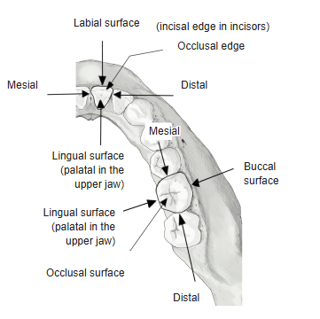
· Labial surface: the surface facing the lips, i.e. the front surface.
· Lingual / palatal surface: the back surface; lingual in the lower jaw, palatal in the upper jaw.
· Occlusal edge: the edge where upper and lower teeth meet; in the incisors it is almost straight and is called the incisal edge.
⟨Molars⟩
· Buccal surface: the surface facing the cheek, i.e. the outer surface.
· Lingual / palatal surface: the inner surface; lingual in the lower jaw, palatal in the upper jaw.
· Occlusal surface: the surface where upper and lower teeth meet; grooves divide it into cusps.
 Surfaces and edges of the teeth
[Incisors]
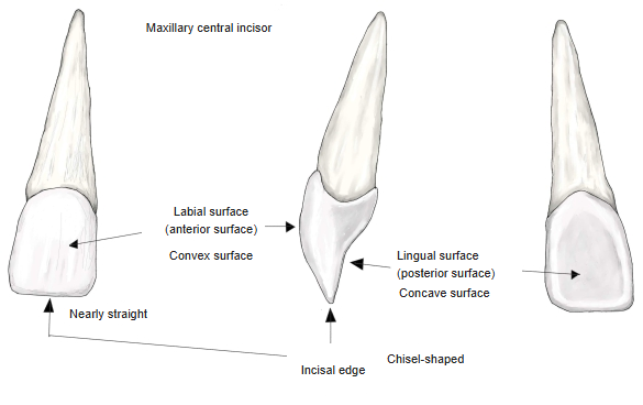
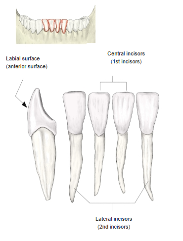
The main features of the incisors:
· The labial (front) surface is convex, the lingual (back) surface slightly concave.
· The free end of the crown is almost straight and is called the "incisal edge".
· The cutting end of the crown is thin and sharp, like a chisel.
· The root is slightly longer than the crown.
· The upper ones are larger than the lower ones, and the upper first incisor (central incisor) is the largest.
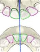 Incisors
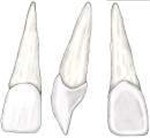 Upper central incisor
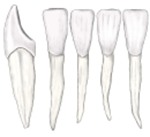 Lower incisor
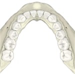 Lower dental arch
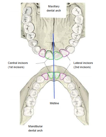
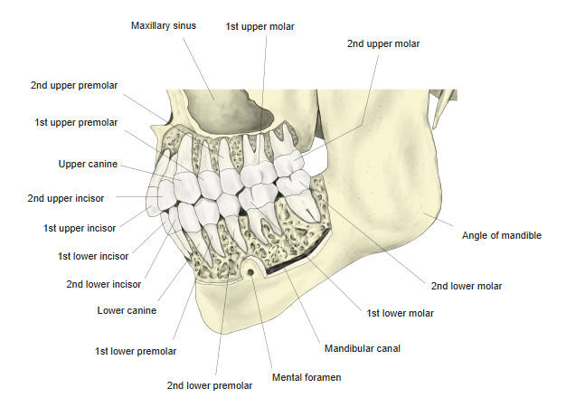
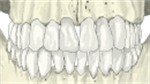 Upper and lower teeth (front)
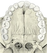 Upper dental arch
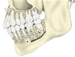 Teeth and alveolar bone
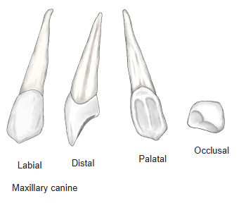
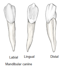
[Canines]
· As in the incisors, the labial surface is convex and the lingual surface concave.
· The crown is pointed like a cone and projects further than the other teeth, so it is suited to biting off and holding food.
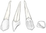 Upper canine
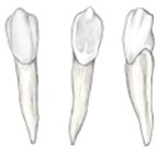 Lower canine
- Lower dental arch
- Upper and lower teeth (front)
- Upper dental arch
- Teeth and alveolar bone
[Premolars]
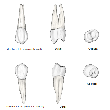
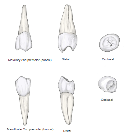
In the lower premolars the root is single and shows only a longitudinal groove on its front and back surfaces; in the upper jaw the apex of the 1st premolar often divides into 2 roots, buccal and lingual.
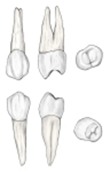 1st premolar
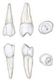 2nd premolar
- Lower dental arch
- Upper and lower teeth (front)
- Upper dental arch
- Teeth and alveolar bone
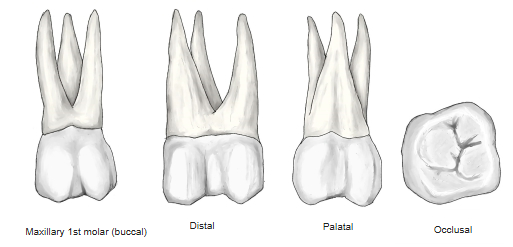
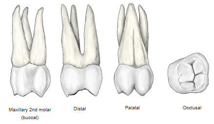
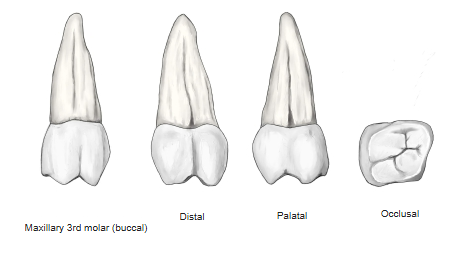
[Molars]
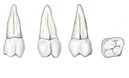
· Cusp of Carabelli: a cusp sometimes found on the lingual (palatal) side of the mesial cusp of the upper 1st molar.
■ Third molars ■
· Formation starts at about 8 years and is complete at about 14 years.
· In general they erupt earlier in men than in women (for the second molars the reverse is common).
· About 25% of people have no third molars (congenital absence).
· In English they are called "wisdom teeth" because they erupt at the age when wisdom comes.
 Upper 1st molar (6 years)
 Upper 2nd molar (12 years)
 Upper 3rd molar
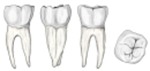 Lower 1st molar (6 years)
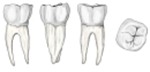 Lower 2nd molar (12 years)
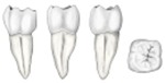 Lower 3rd molar
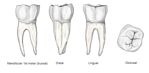
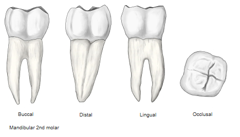
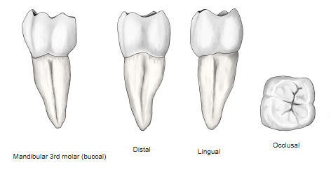
- Upper and lower teeth (front)
- Upper dental arch
- Teeth and alveolar bone
Vessels and nerves
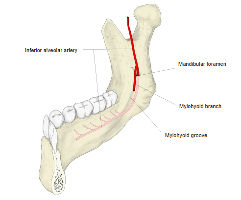
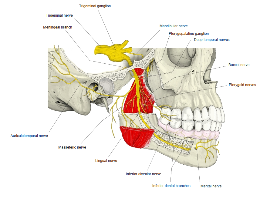
The vessels and nerves supplying the dental pulp and the periodontal ligament are as follows.
| Arteries | Branches of the maxillary artery Lower teeth: dental and gingival branches of the inferior alveolar artery Upper teeth: anterior superior alveolar artery, middle superior alveolar artery (?), posterior superior alveolar artery |
|---|---|
| Veins | Accompany the arteries: inferior alveolar vein, anterior, middle and posterior superior alveolar veins |
| Lymphatics | To the submandibular lymph nodes; part of the lower teeth drains to the submental lymph nodes |
| Nerves | Branches of the trigeminal nerve Lower teeth: inferior alveolar nerve (from the mandibular nerve) Upper teeth: anterior, middle and posterior superior alveolar nerves (from the maxillary nerve) |
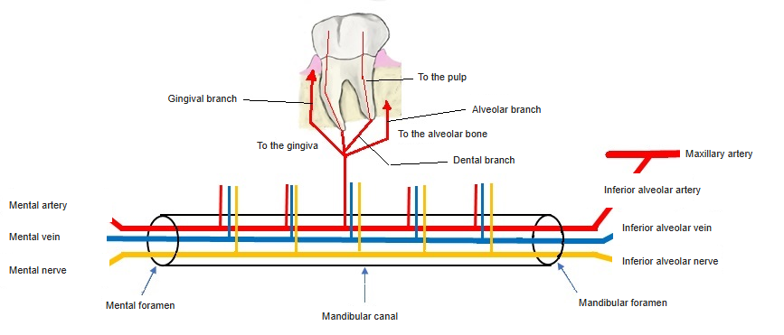
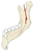 Inferior alveolar artery
 Mandibular nerve
Development
The enamel of the tooth arises from the ectoderm (oral epithelium); the dentin and the cementum arise from the mesoderm.
In the 7th fetal week the epithelium of the jaw margin proliferates and grows into the deeper connective tissue as a ridge: this is the dental lamina.
In the 3rd fetal month, club-shaped thickenings, as many as the deciduous teeth, appear at regular intervals along the free edge of the dental lamina: these are the tooth germs, the first rudiments of the deciduous teeth. Each thickening develops into the enamel organ, which is concerned with forming enamel. At the same time, mesenchymal connective tissue cells gather under the enamel organ and form the dental papilla.
At first the dental lamina is broadly joined to the enamel organ, but it gradually narrows into a thin connecting bridge. Later a second club-shaped thickening appears on the lingual side of the enamel organ of each deciduous tooth, and the germ of the permanent tooth arises there (5th fetal month). As the enamel organ develops, 3 cell layers can be distinguished: the outer layer is the outer enamel epithelium, the inner layer the inner enamel epithelium, and between them lies the enamel pulp (stellate reticulum). The outer enamel epithelium, on the outside of the enamel organ, is a simple cuboidal epithelium. The inner enamel epithelium, a simple columnar epithelium covering the hollow under the enamel organ, differentiates into ameloblasts. The enamel pulp is filled with star-shaped cells.
Along with these changes, the mesodermal cells on the surface of the dental papilla become columnar and line up like an epithelium: these are the odontoblasts. The dental follicle (sac) arises from the connective tissue around the enamel organ. In the 20th fetal week the odontoblasts secrete predentin, a structureless intercellular substance; collagen fibrils gradually appear, the matrix calcifies, and dentin is formed.
Soon after dentin formation begins, enamel formation starts: the ameloblasts, differentiated from the inner enamel epithelium, form pre-enamel where they face the dentin; it calcifies, and the first enamel rods are formed. As development proceeds, the inner enamel epithelium stops forming enamel, leaving a cuticle; this remains as the enamel cuticle covering the surface of the tooth, but it wears away some time after eruption. Cementum arises from the connective tissue surrounding the enamel organ, i.e. the dental follicle, and the periodontal ligament also arises from the same tissue.
The permanent teeth develop in the same way as the deciduous teeth, from a part of the dental lamina deeper than the deciduous teeth, in the 24th fetal week. When the teeth are replaced, the roots of the deciduous teeth are broken down and resorbed by the developing permanent teeth, and their crowns are pushed out of the alveoli.
Related terms
Main anatomical terms related to the teeth, in alphabetical order.
| A | Accessory root, Apex of root, Apical foramen |
| B | Buccal surface |
| C | Canine, Cementum, Clinical crown, Clinical root, Coronal pulp, Crown, Crown cavity, Cusp of tooth |
| D | Deciduous teeth, Dental alveoli, Dental pulp, Dentin, Distal cusp, Distal root, Distobuccal cusp, Distolingual cusp, Distopalatal cusp |
| E | Enamel |
| I | Incisor, Inferior dental arch |
| L | Lingual surface |
| M | Mesial root, Mesiobuccal root, Mesiolingual root, Molar |
| N | Neck of tooth |
| O | Occlusal surface |
| P | Palatal root, Palatal surface, Periodontium, Permanent teeth, Premolar, Pulp cavity |
| R | Radicular pulp, Root canal, Root of tooth |
| S | Superior dental arch |
| V | Vestibular surface |
| W | Wisdom tooth (= third molar) |
References and sources
- Further reading: Tooth, Wikipedia.