Abdominal wall (Latin: paries abdominis)
- Overview
- Each wall in detail
- Reference websites
- (Wikipedia, websites with illustrations)
- Related terms
Overview
· It is usually divided into an anterior, a lateral and a posterior wall. The anterior and lateral walls are mostly muscle; the posterior wall also contains the lumbar vertebrae and is much thicker. Sources differ on which structures count as part of the abdominal wall; in clinical texts the term often means only the anterolateral wall.
of the
abdominal
cavity
- Upper wall (roof) : the diaphragm (a sheet of muscle), the boundary between the thoracic cavity and the abdominal cavity.
- Front, side and back walls (the abdominal wall) : from outside: skin, subcutaneous tissue, the muscle layer (the main part), the skeleton and the peritoneum.
- Lower wall (floor) : the pelvic brim (terminal line) of the pelvis, the line joining the promontory, arcuate line, pecten pubis and the upper edge of the pubic symphysis.
The details differ a little from region to region, but the basic make-up of the abdominal wall is shown below. From outside to inside the basic layers are: skin, subcutaneous tissue, muscles with their fasciae, the transversalis fascia, extraperitoneal fat and the parietal peritoneum.
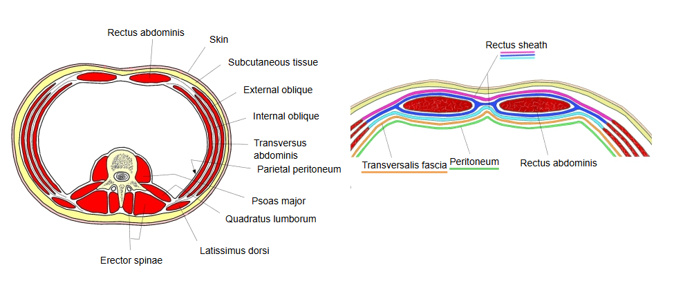
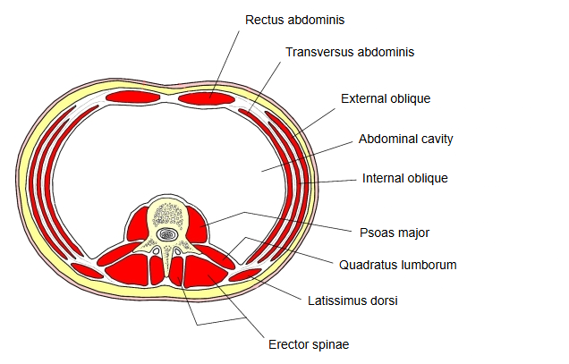
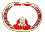 Transverse section of the abdominal cavity
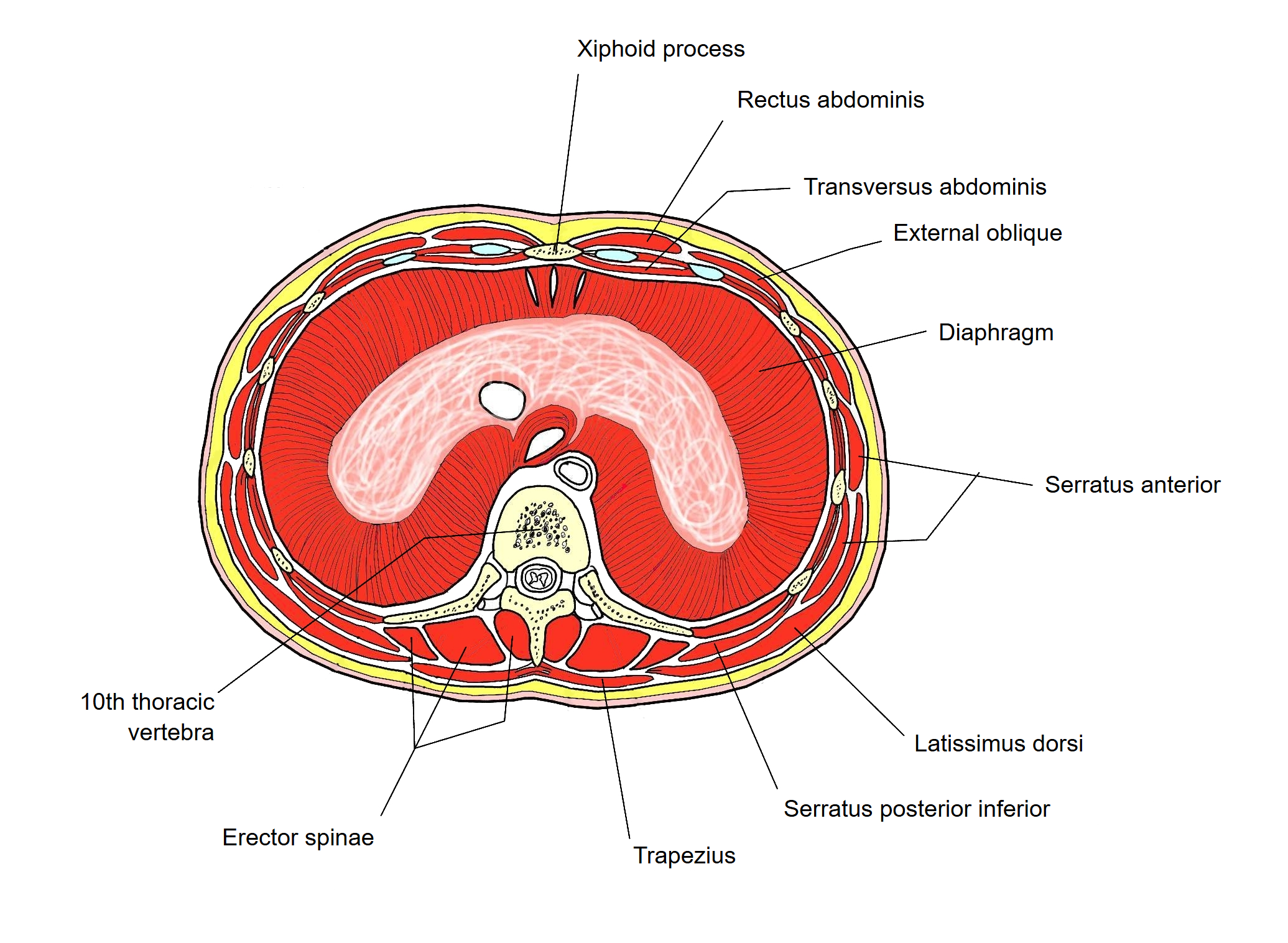
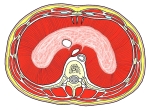 Transverse section at the tenth thoracic vertebra (upper abdomen)
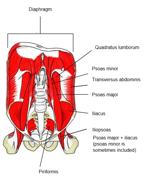
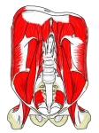 Abdominal cavity (side and back walls)
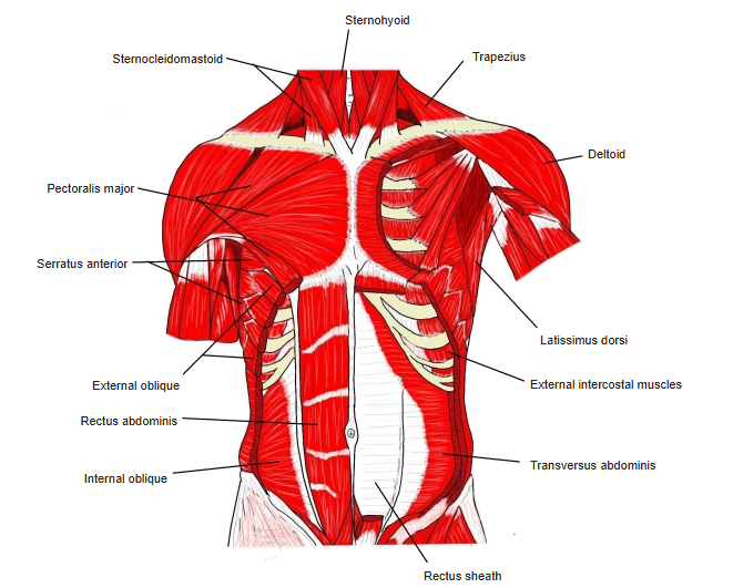 Chest to abdomen (muscles)
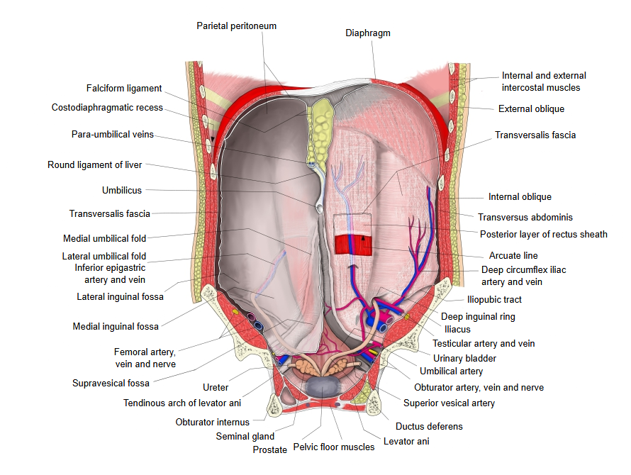
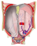 Anterior abdominal wall seen from behind
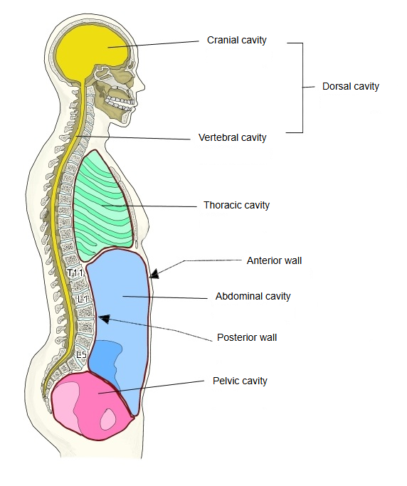
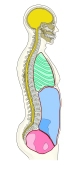 Body cavities
Each wall in detail
Anterior wall
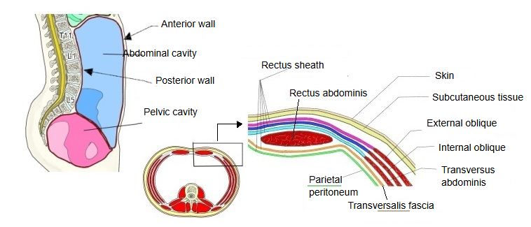
From outside: skin → subcutaneous tissue → fascia → muscle → transversalis fascia and extraperitoneal fat → peritoneum (parietal peritoneum)
Skin: from outside, the epidermis and the dermis.
Subcutaneous tissue: mainly adipose tissue, with blood vessels and nerves running through it and thin connective tissue layers spread within it.
Muscles: the rectus abdominis in the rectus sheath, and the front parts of the flat lateral muscles (external oblique, internal oblique, transversus abdominis).
Bone: only a small contribution, from the lower sternum and some of the costal cartilages.
Membranes: the fasciae of the muscles; on the inner side, the transversalis fascia and the parietal peritoneum.
Posterior wall
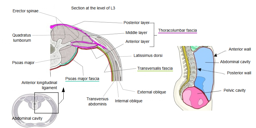
The posterior wall of the abdominal cavity is thick and strong and protects the organs more than the anterior wall does.
Leaving aside the outermost layers such as the skin, it is made up of:
Bone: from about the eleventh thoracic vertebra above to the fifth lumbar vertebra
(sometimes also the upper sacrum and the ilium).
Muscles: the deep (intrinsic) back muscles, chiefly the erector spinae; the iliopsoas (psoas major and iliacus), the quadratus lumborum and the crura of the diaphragm.
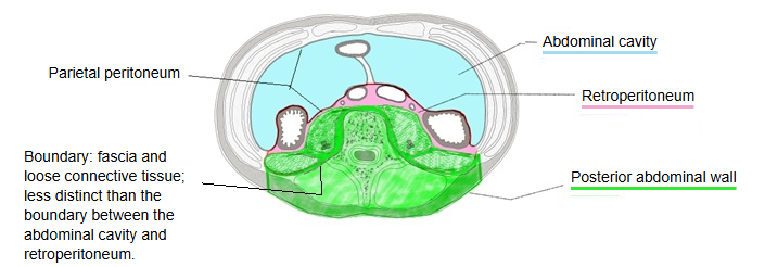
Membranes: the fascia of each muscle, the thoracolumbar fascia (three layers) and, innermost, the parietal peritoneum.
The transversalis fascia is sometimes included as well.
· The kidneys, suprarenal glands, aorta and inferior vena cava lie on this wall behind the parietal peritoneum, in the retroperitoneum.
Lateral wall
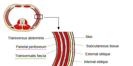
Leaving aside the outermost layers such as the skin, it is made up of:
Bone: the lower ribs above and the iliac crest of the ilium below also take part.
Muscles: from outside inward, the external oblique, the internal oblique and the transversus abdominis.
Membranes: the fascia of each muscle, the transversalis fascia and, innermost, the parietal peritoneum.
What about the serratus anterior, the latissimus dorsi and the internal and external intercostal muscles?
They lie nearby, but are usually not counted as parts of the abdominal wall.
Reference websites
[Wikipedia]
· Abdominal wall, Wikipedia.
[Websites with illustrations]
· Vessels on the posterior abdominal wall (figure) (scuguide.com)

· The anterolateral abdominal wall: muscles (TeachMeAnatomy)
· Abdominal wall: layers, muscles and fascia (Kenhub)
Related terms
- Rectus abdominis
- Transversus abdominis
- Internal oblique
- Quadratus lumborum
- Transversalis fascia
- Parietal peritoneum
- Peritoneal cavity
References and sources
- Further reading: Abdominal wall, Wikipedia.
- Federative International Programme for Anatomical Terminology (FIPAT). Terminologia Anatomica, 2nd ed. (TA2), 2019.