Latin: os coxae · also called the innominate bone or coxal bone
Overview
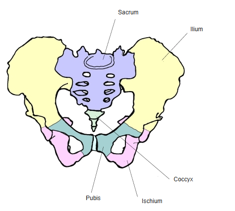
- The hip bone is a large, flat bone made up of three parts: the ilium, the ischium and the pubis.
- The left and right hip bones, together with the sacrum and coccyx, form the pelvis; the hip bones lie on either side of the sacrum and form the front and side walls of the pelvis.
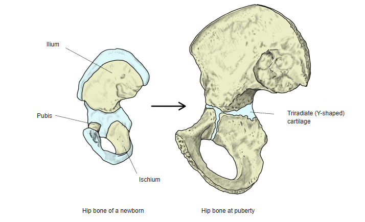
- Until puberty the three parts are joined by a Y-shaped (triradiate) cartilage in the acetabulum; after puberty this cartilage is replaced by bone and they become one complete bone. The three parts meet at the acetabulum.
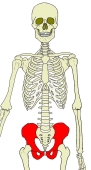
Position of the hip bone 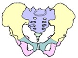
Pelvis (anterior) 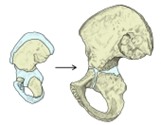
Ossification
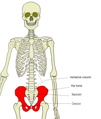
- Bones of the lower limb (one side)
- Pelvic girdle
- Hip bone ← ilium, ischium and pubis fused
- Free lower limb
- Thigh and knee
- Femur
- Patella
- Leg
- Tibia ← big-toe side
- Fibula ← little-toe side
- Foot
- Tarsal bones (7)
- Metatarsal bones (5)
- Phalanges (14)
- Thigh and knee
- Pelvic girdle
Illustrations
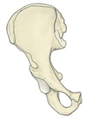
Right hip bone (anterior) 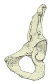
Right hip bone (anterior oblique) 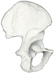
Right hip bone (lateral) 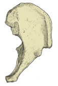
Left hip bone (from above) 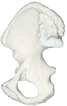
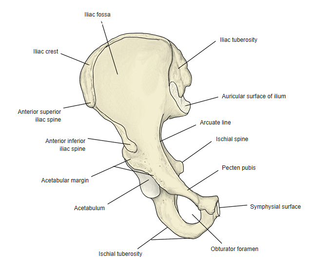
Right hip bone (medial)
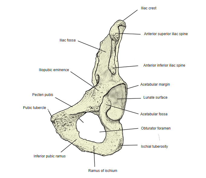
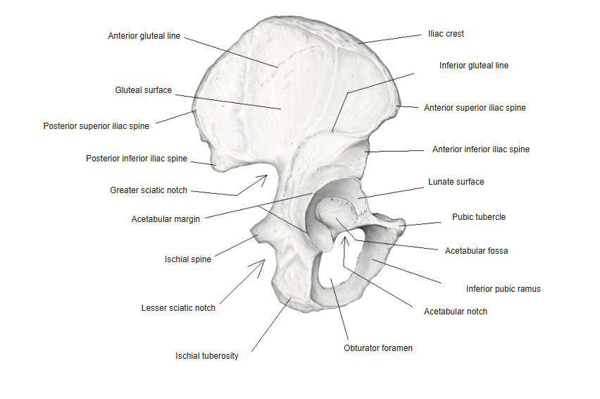
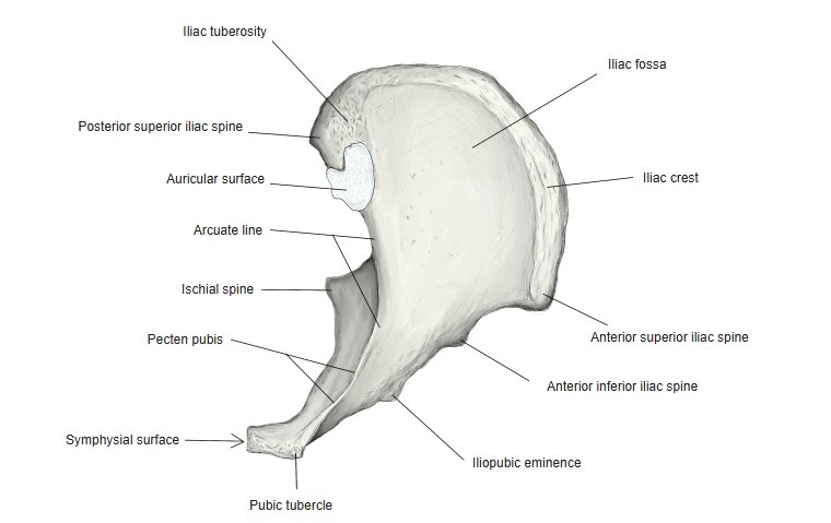
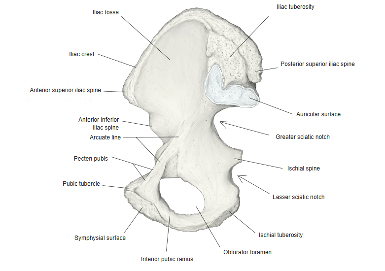
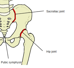
Articulations
| # | Bone | Joint |
|---|---|---|
| 1 | Femur | Hip joint |
| 2 | Sacrum | Sacroiliac joint |
| 3 | Hip bone of the other side | Pubic symphysis |
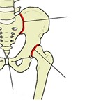
Joints of the hip bone
Ligaments
The following ligaments attach to the hip bone. The list is intended to be comprehensive but may omit minor ligaments.
| # | Ligament | Connects |
|---|---|---|
| 1 | Anterior sacroiliac ligament | Sacroiliac joint (ilium – sacrum) |
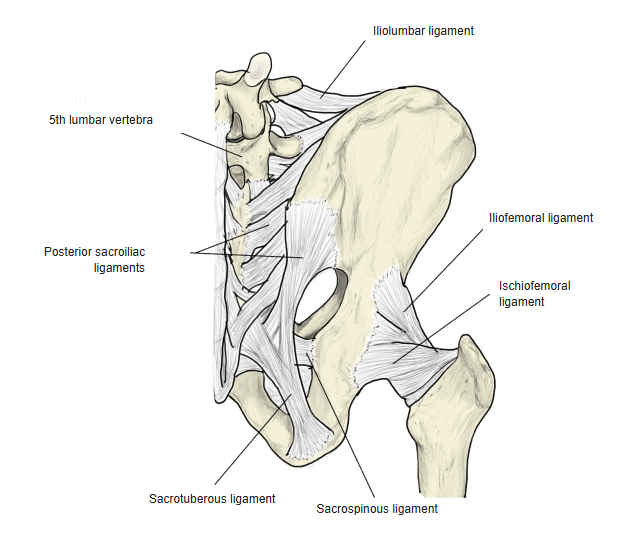
| 2 | Posterior sacroiliac ligament | Sacroiliac joint (ilium – sacrum); divided into short and long parts |
| 3 | Interosseous sacroiliac ligament | Sacroiliac joint (ilium – sacrum) |
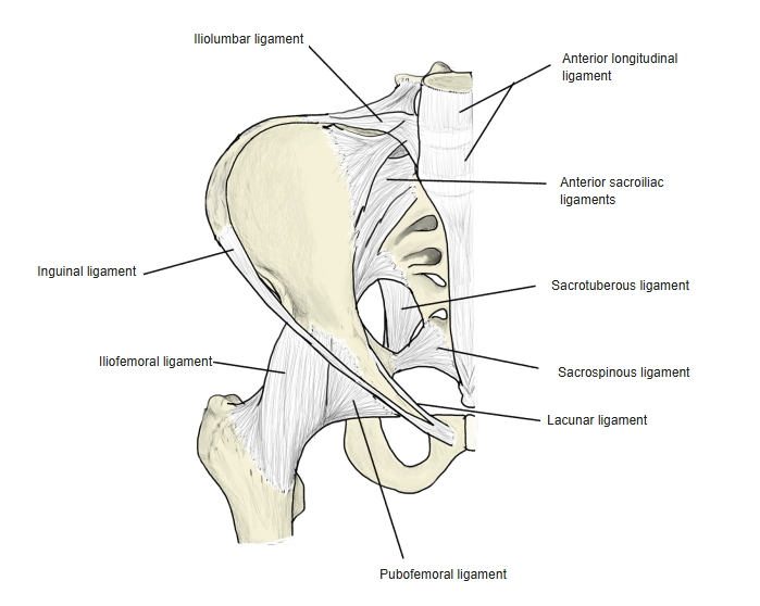
| 4 | Iliofemoral ligament | Hip joint (hip bone – femur) |
| 5 | Pubofemoral ligament | Hip joint (hip bone – femur) |
| 6 | Ischiofemoral ligament | Hip joint (hip bone – femur) |
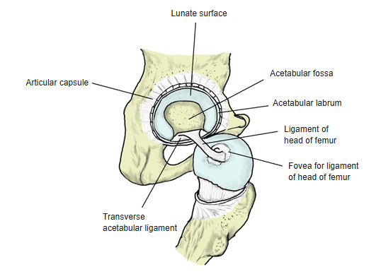
| 7 | Ligament of head of femur | Hip joint (hip bone – femur) |
| 8 | Transverse acetabular ligament | Hip joint (hip bone – femur) |
| 9 | Zona orbicularis | Hip joint (hip bone – femur) |
| 10 | Superior pubic ligament | Pubic symphysis |
| 11 | Inferior pubic ligament (arcuate pubic ligament) | Pubic symphysis |
| 12 | Iliolumbar ligament | Ilium – lumbar vertebrae |
| 13 | Pectineal ligament | Pecten pubis |
| 14 | Inguinal ligament | Anterior superior iliac spine – pubic tubercle |
| 15 | Lacunar ligament | Inguinal ligament – pecten pubis |
| 16 | Sacrospinous ligament | Hip bone – sacrum and coccyx |
| 17 | Sacrotuberous ligament | Hip bone – sacrum and coccyx |
| 18 | Puboprostatic ligament | Pubis – prostate (male) |
| 19 | Pubovesical ligament | Pubis – bladder (female) |
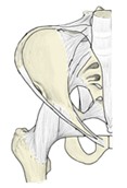
Ligaments of the pelvis (anterior) 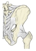
Ligaments of the pelvis (posterior) 
Hip joint (acetabulum and head of femur)
Muscle attachments
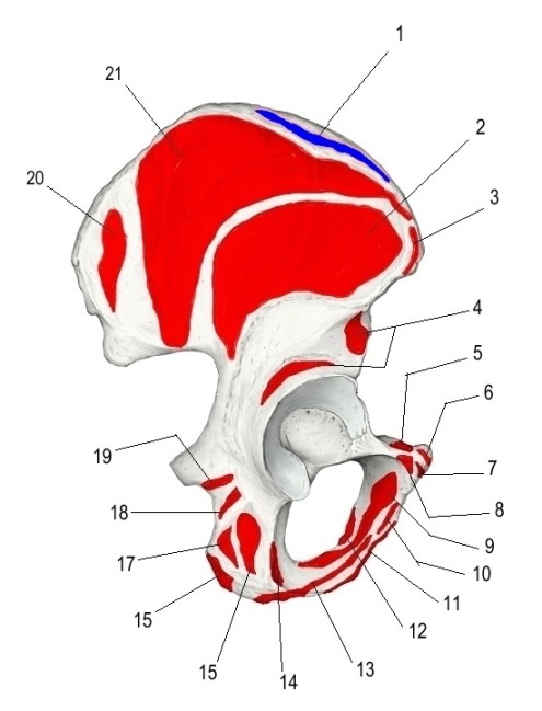
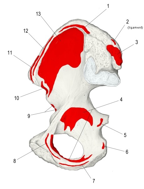
- Origin of: Obturator externus, Gemellus inferior, Levator ani, Ischiocavernosus, Gemellus superior, Gluteus minimus, Adductor minimus, Pyramidalis, Erector spinae, Superficial transverse perineal, Tensor fasciae latae, Rectus femoris, Biceps femoris (long head), Quadratus femoris, Gluteus maximus, Adductor magnus, Adductor brevis, Pectineus, Gluteus medius, Iliacus, Adductor longus, Internal oblique, Obturator internus, Sphincter urethrae, Gracilis, Semitendinosus, Semimembranosus, Coccygeus, Transversus abdominis, Rectus abdominis, Sartorius, Quadratus lumborum, Latissimus dorsi
- Insertion of: External oblique, Psoas minor
Red: origin · blue: insertion in the figures below.
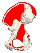
Muscle attachments (medial surface) 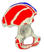
Muscle attachments (lateral surface) 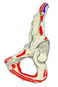
Muscle attachments (anterior oblique) 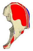
Muscle attachments (from above)
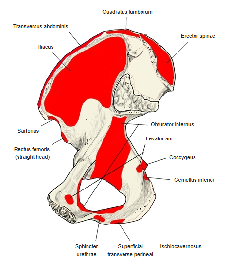
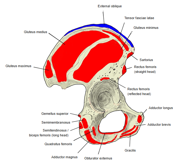
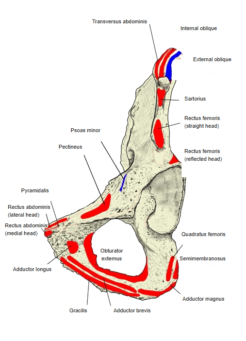
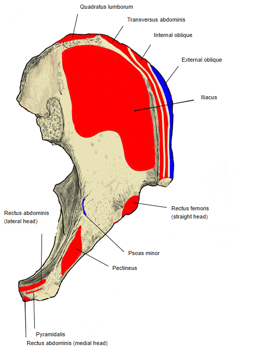
Numbered figures with the names of the muscles:
Right hip bone (lateral view)
Right hip bone (medial view)
Parts of the ilium
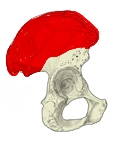
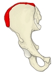
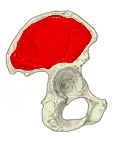
- Ala of ilium – the upper, wing-like part of the ilium; its upper border is the iliac crest. The boundary with the body of the ilium is not distinct.
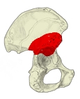
- Body of ilium – the lower part of the ilium, forming part of the acetabulum.
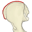
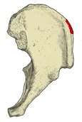
- Iliac crest – the thickened upper border of the ala; it is thinner in its middle part.
- Outer lip – the outer edge of the iliac crest. Insertion: external oblique.
- Intermediate line – the ridge on top of the iliac crest between the outer and inner lips. Origin: internal oblique.
- Inner lip – the inner edge of the iliac crest. Origin: transversus abdominis.
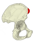
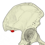
- Tubercle of iliac crest – a palpable bump on the outer lip, about 5 cm behind the anterior superior iliac spine, where the anterior gluteal line meets the crest.
- Anterior superior iliac spine – the prominent front end of the iliac crest; easy to feel under the skin. Origin: tensor fasciae latae, sartorius.
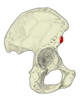
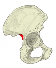
- Anterior inferior iliac spine – the projection below the anterior superior iliac spine.
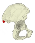
- Posterior superior iliac spine – the projection at the back end of the iliac crest.
- Posterior inferior iliac spine – the projection below the posterior superior iliac spine.
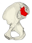
- Iliac fossa – the shallow, smooth hollow forming about the front two-thirds of the inner surface of the ala. Origin: iliacus.
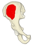
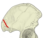
- Gluteal surface – the outer surface of the ilium, where the gluteal muscles attach; its front part bulges slightly and its back part is slightly hollow.
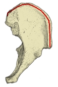
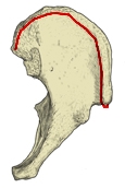
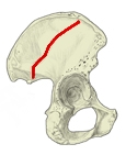
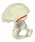
- Anterior gluteal line – the longest of the three gluteal lines, running across the middle of the gluteal surface from front-above to back-below; it separates the origins of gluteus medius and gluteus minimus.
- Posterior gluteal line – a short line behind the anterior one, running almost straight down from the iliac crest; it separates gluteus maximus from gluteus medius.
- Inferior gluteal line – the gluteal line running obliquely just above the acetabulum.
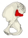
- Auricular surface – the ear-shaped, cartilage-covered surface that articulates with the sacrum at the sacroiliac joint, which allows almost no movement.
- Iliac tuberosity – the rough area just above the auricular surface; the interosseous sacroiliac ligament attaches here.
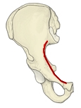
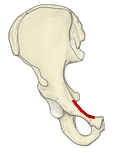
- Arcuate line – the ridge on the inner surface running forwards from the lower end of the auricular surface; it continues into the pecten pubis.
- Greater sciatic notch – the large notch on the posterior border, above and behind the acetabulum.
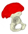
Ala of ilium 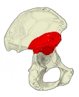
Body of ilium 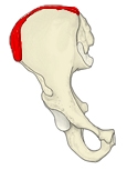
Iliac crest 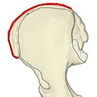
Outer lip 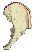
Intermediate line 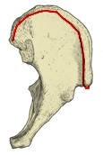
Inner lip 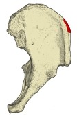
Tubercle of iliac crest 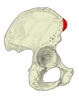
Anterior superior iliac spine 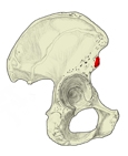
Anterior inferior iliac spine 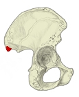
Posterior superior iliac spine 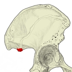
Posterior inferior iliac spine 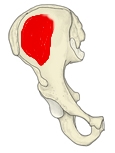
Iliac fossa 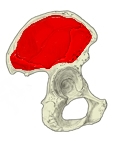
Gluteal surface 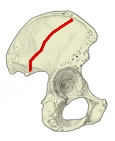
Anterior gluteal line 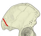
Posterior gluteal line 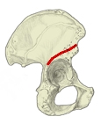
Inferior gluteal line 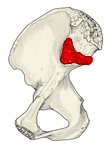
Auricular surface 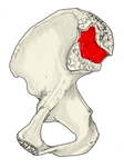
Iliac tuberosity 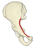
Arcuate line 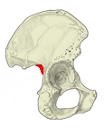
Greater sciatic notch
Parts of the ischium
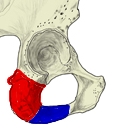
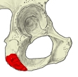
- Body of ischium – the part of the ischium that forms part of the acetabulum, above the ischial spine.
- Ramus of ischium – the rest of the ischium; it runs from the body past the ischial tuberosity and joins the inferior pubic ramus.
- Ischial tuberosity – the very rough area on the back of the ischium, origin of many muscles: adductor magnus, biceps femoris (long head), semitendinosus, semimembranosus and quadratus femoris. The sacrotuberous ligament also attaches here.
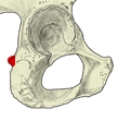
- Ischial spine – the triangular projection on the upper part of the posterior border. Origin: gemellus superior, coccygeus. The sacrospinous ligament attaches here.
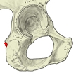
- Lesser sciatic notch – the notch just below the ischial spine.
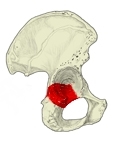
Body of ischium 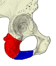
Ramus of ischium 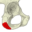
Ischial tuberosity 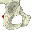
Ischial spine 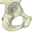
Lesser sciatic notch
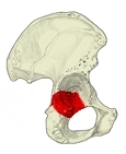
Parts of the pubis
- Body of pubis – the wide, flattened main part of the pubis; it bears the symphysial surface medially and forms part of the acetabulum laterally.
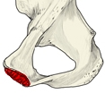
- Superior pubic ramus – the part projecting forwards from the body of the pubis; it carries the pecten pubis, the iliopubic eminence, the pubic tubercle and the obturator groove.
- Inferior pubic ramus – the part running down and laterally from the front end of the superior ramus, joining the ramus of the ischium.
- Symphysial surface – the oval, uneven surface where the two pubic bones meet at the pubic symphysis. Its relief changes with age, so it is used to estimate age at death.
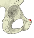
- Pubic tubercle – the small projection on the upper front end of the pubis.
- Pecten pubis – the sharp upper edge of the superior pubic ramus, forming part of the line that continues from the arcuate line.
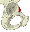
- Iliopubic eminence – the elevation at the upper end of the superior pubic ramus, at the front edge of the acetabulum.
- Obturator crest – a ridge on the superior pubic ramus at the upper front edge of the obturator foramen; it runs from the pubic tubercle to the acetabular notch.
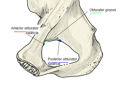
- Obturator groove – the groove on the superior pubic ramus at the upper front corner of the obturator foramen, between the anterior and posterior obturator tubercles.
- Anterior and posterior obturator tubercles – small tubercles at either end of the obturator groove.
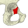
Body of pubis 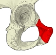
Superior pubic ramus 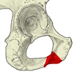
Inferior pubic ramus 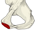
Symphysial surface 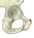
Pubic tubercle 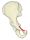
Pecten pubis 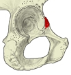
Iliopubic eminence 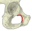
Obturator crest 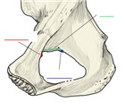
Obturator groove and obturator tubercles
Acetabulum and obturator foramen
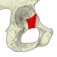
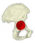
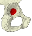
- Acetabulum – the deep, cup-shaped hollow on the outer side, above the obturator foramen, where all three parts of the hip bone meet. It receives the head of the femur to form the hip joint; a fibrocartilaginous acetabular labrum around it makes it deeper.
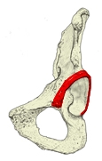
- Acetabular margin – the raised rim around the acetabulum; it is interrupted by the acetabular notch, so it does not go all the way round.
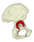
- Lunate surface – the crescent-shaped, cartilage-covered articular surface of the acetabulum.
- Acetabular fossa – the deep central part of the acetabulum that is not covered with cartilage.
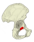
- Acetabular notch – the gap in the acetabular margin; the lower end of the acetabular fossa forms its floor.
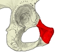
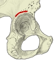
- Supra-acetabular groove – the groove above the acetabular margin. Origin: rectus femoris (reflected head).
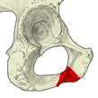
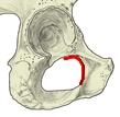
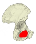
- Obturator foramen – the large opening below the acetabulum; in life it is almost completely closed by the obturator membrane.
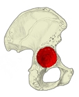
Acetabulum 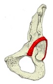
Acetabular margin 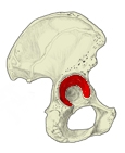
Lunate surface 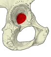
Acetabular fossa 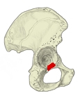
Acetabular notch 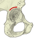
Supra-acetabular groove 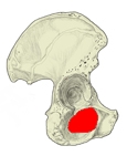
Obturator foramen
References and sources
- Further reading: Hip bone, Wikipedia.