Hip joint (Latin: articulatio coxae)
Also called the coxal joint
- Overview
- Muscles
- Ligaments
- Axes of movement
- Videos
- Angles of the femur
- Where it is found
- Related terms
Overview
The hip joint, at the root of the lower limb, is one of the joints of the bones of the lower limb. It is formed by the femur (its head) and the hip bone (its acetabulum), and joins the free lower limb to the pelvic girdle. It is one of the largest joints of the body.
of the
lower limb
- Joints of the pelvic girdle : union of the parts of the hip bone; sacroiliac joint (hip bone with the sacrum); ligaments joining the hip bone to the sacrum and coccyx (sacrotuberous and sacrospinous ligaments); pubic symphysis.
- Joints of the free lower limb
- Hip joint : head of the femur ⇔ acetabulum of the hip bone: one of the largest joints of the body.
- Knee joint : femur, tibia and patella.
- Tibiofibular joints : between the tibia and the fibula.
- Joints of the foot : ankle joint and the joints of the foot bones.
It is a ball-and-socket joint of the deep, cup-shaped type (an enarthrosis). More than half of the head of the femur, the articular head, sits inside the acetabulum, the articular socket. This deep fit, together with very strong ligaments, makes it far more stable than the shoulder joint, another ball-and-socket joint, but its movements are much more limited.
joint
- Articular head : ⇒ head of femur: more than half of it sits in the acetabulum ⇒ enarthrosis; its movements are much more limited than those of the shoulder joint (ball-and-socket joint).
- Articular socket : ⇒ acetabulum
 Right lower limb (anterior)
 Right lower limb (posterior)
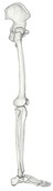 Right lower limb (lateral)
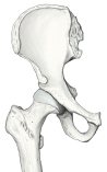 Right hip joint (anterior)
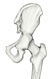 Right hip joint (posterior)
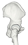 Right hip joint (lateral)
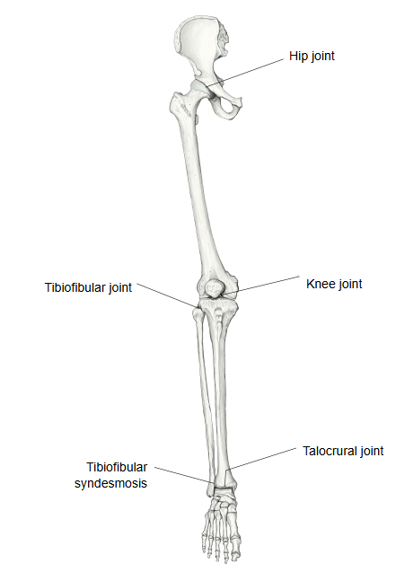
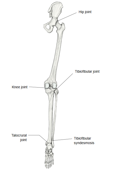
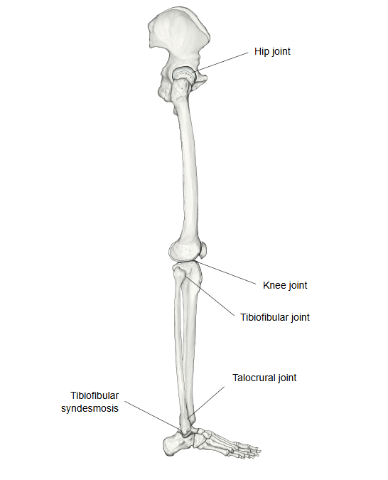
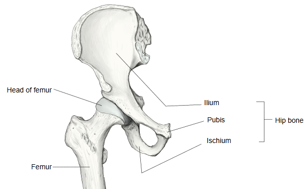
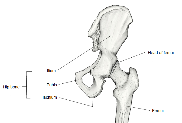
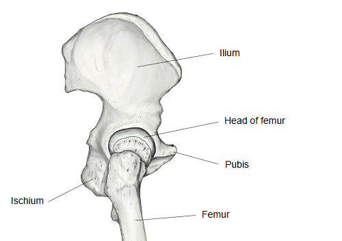
Looking more closely at the parts of the joint:
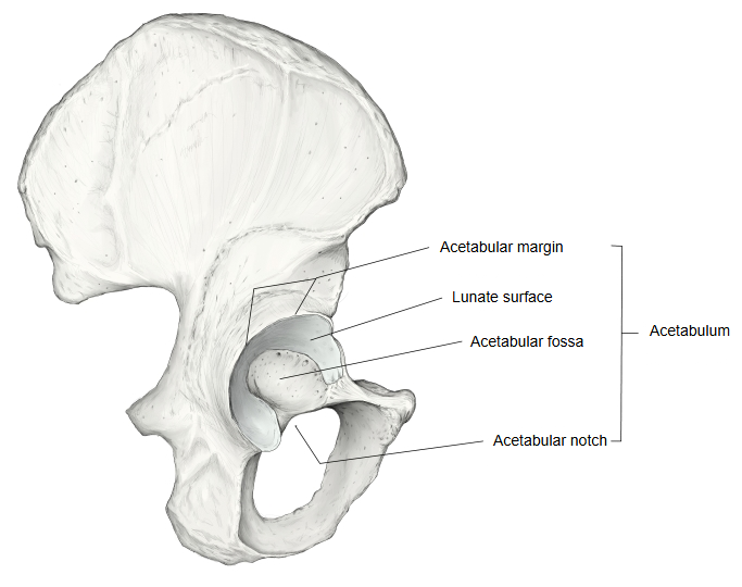
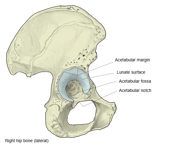
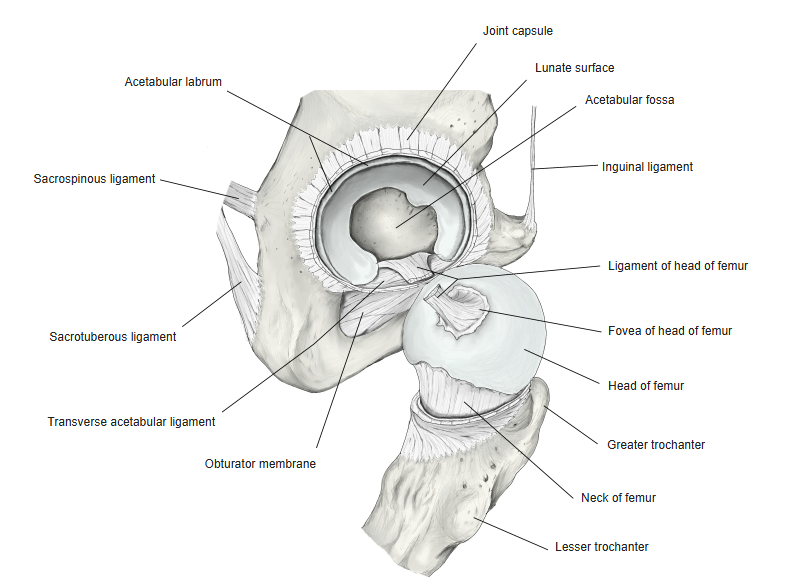
Acetabulum
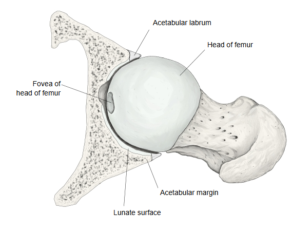
| Lunate surface | The horseshoe-shaped surface (articular surface) that meets the head of the femur; covered with cartilage. It is named for its half-moon shape. |
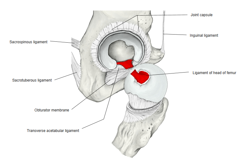
| Acetabular fossa | The central part of the acetabulum without cartilage. It is filled with a fat pad (adipose tissue) covered by synovial membrane, and the ligament of head of femur arises here. |
| Acetabular labrum | A ring of fibrocartilage running all the way round the acetabular margin. It deepens the socket and helps to keep the acetabulum and the femur stable; it bridges the acetabular notch as the transverse acetabular ligament. |
 Right hip bone (lateral)
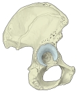 Acetabulum (lateral)
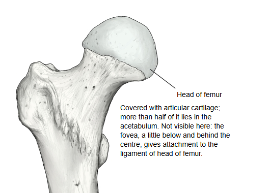
Head of femur
Covered with articular cartilage. A little below and behind its top is a small pit, the fovea of the head of femur, without cartilage, where the ligament of head of femur is attached.
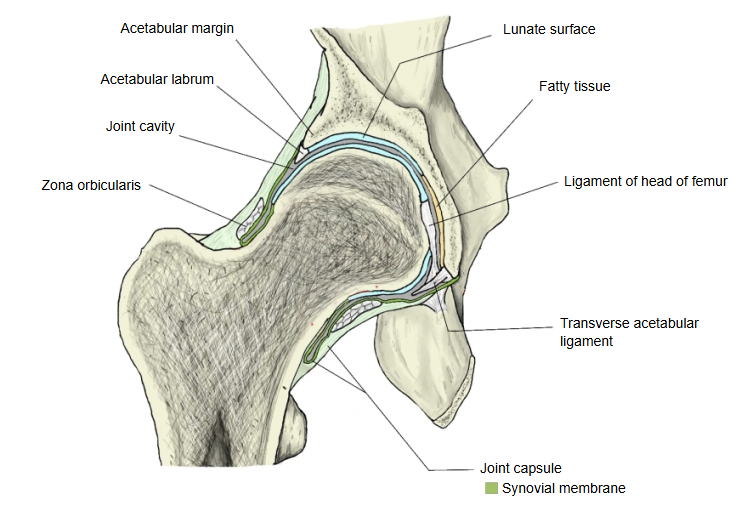
Joint capsule
The joint capsule is strong and dense. Its attachments are as follows.
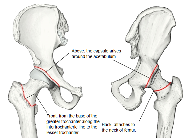
Above, the joint capsule arises around the acetabulum, just outside the labrum. Below, in front it is attached along the intertrochanteric line, from the base of the greater trochanter to the lesser trochanter; behind it is attached higher, to the neck of femur, about a finger's breadth above the intertrochanteric crest, so much of the back of the neck lies outside the capsule. At both ends the line of attachment reaches the greater and lesser trochanters.
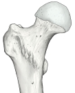 Head of femur
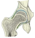 Hip joint (coronal section)
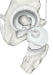 Transverse acetabular ligament
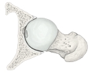 Acetabulum and head of femur (section)
Muscles
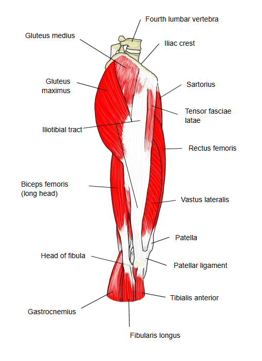
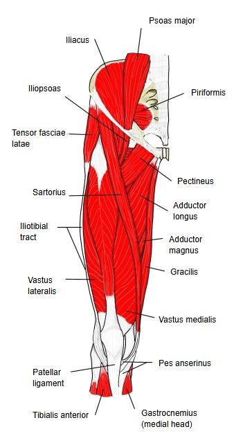
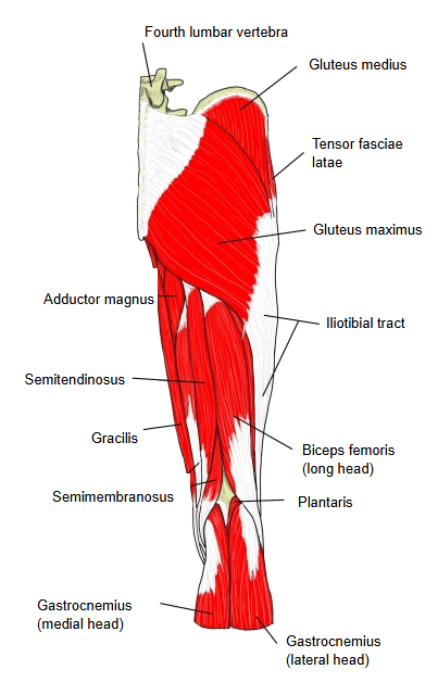
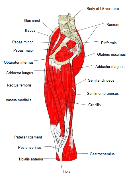
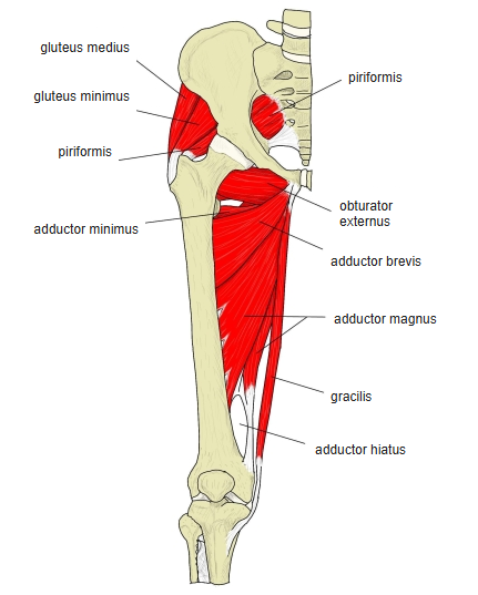
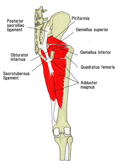
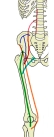
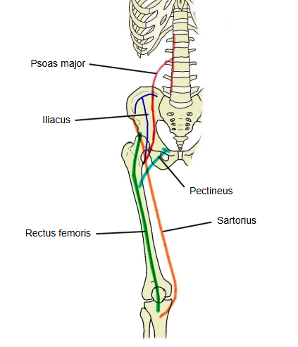
The following are the muscles that act on the hip joint, divided into four groups by their position.
| Front | 1 | psoas major | m. psoas major |
| 2 | iliacus | m. iliacus | |
| 3 | rectus femoris | m. rectus femoris | |
| 4 | sartorius | m. sartorius | |
| Back | 1 | obturator internus | m. obturatorius internus |
| 2 | obturator externus | m. obturatorius externus | |
| 3 | gemellus superior | m. gemellus superior | |
| 4 | gemellus inferior | m. gemellus inferior | |
| 5 | piriformis | m. piriformis | |
| 6 | quadratus femoris | m. quadratus femoris | |
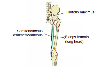
| 7 | gluteus maximus | m. gluteus maximus | |
| 8 | gluteus medius | m. gluteus medius | |
| 9 | gluteus minimus | m. gluteus minimus | |
| 10 | semimembranosus | m. semimembranosus | |
| 11 | semitendinosus | m. semitendinosus | |
| 12 | biceps femoris (long head) | m. biceps femoris, caput longum | |
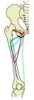
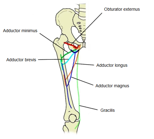
| Medial | 1 | pectineus | m. pectineus |
| 2 | adductor longus | m. adductor longus | |
| 3 | adductor brevis | m. adductor brevis | |
| 4 | adductor magnus | m. adductor magnus | |
| 5 | gracilis | m. gracilis | |
| Lateral | 1 | tensor fasciae latae | m. tensor fasciae latae |
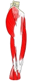 Muscles of the right thigh (lateral)
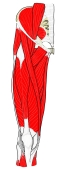 Muscles of the right thigh (anterior)
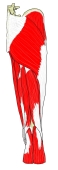 Muscles of the right thigh (posterior)
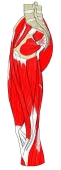 Muscles of the right thigh (medial)
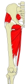 Deep muscles (anterior)
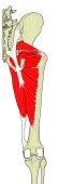 Deep muscles (posterior)
Ligaments
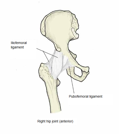
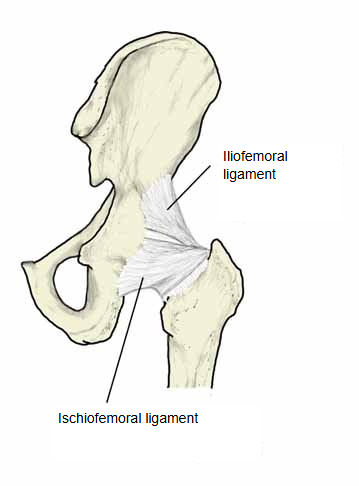
The following are the ligaments that strengthen the hip joint.
| 1 | Ligament of head of femur | A ligament inside the joint capsule, from the acetabular notch and acetabular fossa to the fovea of the head of femur. It carries a small artery, the acetabular branch of the posterior branch of the obturator artery. |
| 2 | Transverse acetabular ligament | Bridges the acetabular notch; together with the acetabular labrum it deepens the socket. |
| 3 | Iliofemoral ligament | The strongest ligament of the body; shaped like an inverted Y, so it is also called the Y ligament. Limits: extension (both bands), lateral rotation and adduction (lateral band), medial rotation (medial band). |
| 4 | Pubofemoral ligament | Runs from the pubic part of the acetabular rim to the lower capsule near the lesser trochanter. Limits mainly abduction (also extension and lateral rotation). |
| 5 | Ischiofemoral ligament | Runs from the ischial part of the acetabular rim to the zona orbicularis and the trochanteric fossa. Limits mainly medial rotation (also adduction with the hip flexed). |
| 6 | Zona orbicularis | Circular fibres of the capsule that encircle the neck of femur. |
 Ligaments (anterior)
 Ligaments (posterior)
- Transverse acetabular ligament
Axes of movement
The axes of movement of the hip joint can be described as follows.
Because the hip joint is a ball-and-socket joint, it has three axes of movement. All three pass through the centre of the head of the femur (the pivot of the joint) and are at right angles to one another. The joint can therefore move in three dimensions, in six main directions.
of the
hip joint
- Transverse axis : flexion and extension
- Sagittal axis : adduction and abduction
- Longitudinal axis : medial rotation and lateral rotation
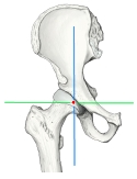 Axes of movement (anterior)
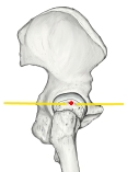 Axes of movement (lateral)
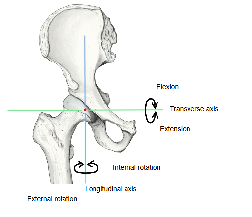
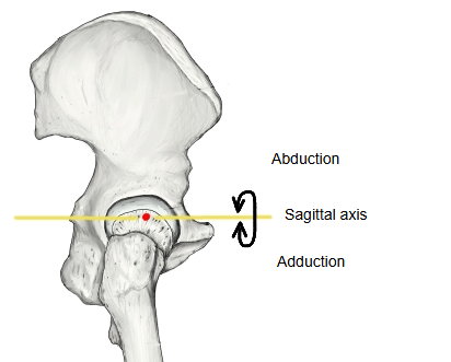
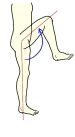
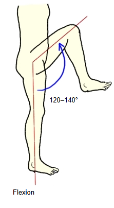
Flexion
The movement of lifting the knee; it is an important movement in walking.
Range of movement: 120°–140° with the knee bent.
 Flexion
 Flexors of the hip
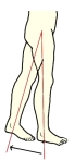
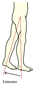
Extension
Swinging the leg backwards is hardly used in daily life, so the range of movement is naturally small.
Range of movement: 15°–20° with the knee straight.
With the knee bent the range is smaller still.
 Extension
 Extensors of the hip
Adduction
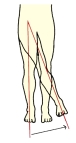
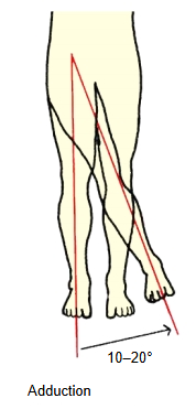
Moving the leg towards and across the midline; it is rarely used in daily life, so its range is quite small.
Range of movement: 10°–20°.
 Adduction
 Adductors of the hip
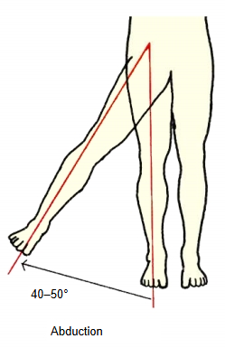
Abduction
Its range is much larger than that of adduction. It is the movement of spreading the legs apart, needed for example in ballet and in the kicks of martial arts.
Range of movement: 40°–50°.
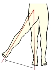 Abduction
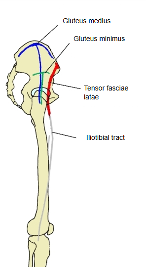
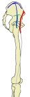 Abductors of the hip
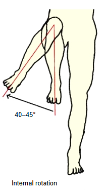
Medial rotation
Turning the leg inwards; it is hardly used on purpose in daily life, and rarely trained deliberately in exercise.
Range of movement: 40°–45°.
No muscle acts only as a medial rotator; gluteus minimus (with the front fibres of gluteus medius) is the prime mover.
Lateral rotation
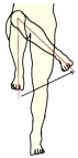
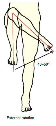
Turning the leg outwards; this movement is needed, for example, in the standing positions of ballet.
Range of movement: 40°–50°.
 Lateral rotation
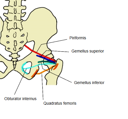
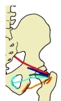 Lateral rotators of the hip
Videos
Angles of the femur
The following terms are related to the hip joint.
Angle of inclination (neck–shaft angle): the angle between the shaft and the neck of the femur. It becomes smaller with growth: newborn 140°–160°; 10 years 130°–140°; adult 125°–135°.
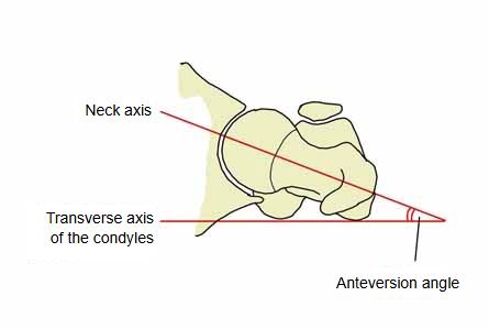
Angle of anteversion (torsion angle): the axis of the neck of the femur is twisted forwards relative to the frontal plane of the shaft (the transverse axis of the condyles); this twist is the angle of anteversion. It becomes smaller with growth: newborn 15°–57°; 1–3 years 20°–50°; adult 12°–15°.
Where it is found
Related terms
- Head of femur
- Acetabulum
- Fovea of head of femur
- Neck of femur
- Hip bone
- Greater trochanter
- Knee joint
References and sources
- Further reading: Hip, Wikipedia.
- Further reading: Iliofemoral ligament, Wikipedia.
- Federative International Programme for Anatomical Terminology (FIPAT). Terminologia Anatomica, 2nd ed. (TA2), 2019.