Vertebral column: related terms
Vertebral column: Overview | Features of the vertebrae | Related terms | Ligaments and membranes | Joints
Terms related to the vertebral column
Accessory process · Ala of sacrum · Anterior arch of atlas · Anterior sacral foramina · Anterior tubercle of atlas · Apex of sacrum · Atlas · Auricular surface of sacrum · Axis · Base of sacrum · Coccygeal cornu · Costal facets · Costal process · Dens · Facet for dens · Groove for vertebral artery · Inferior articular facet · Inferior articular facet of atlas · Inferior articular process · Inferior costal facet · Intermediate sacral crest · Intervertebral foramen · Lamina of vertebral arch · Lateral mass of atlas · Lateral sacral crest · Mammillary process · Medial sacral crest · Median sacral crest · Pedicle of vertebral arch · Posterior arch of atlas · Posterior sacral foramina · Posterior tubercle of atlas · Promontory · Sacral canal · Sacral cornu · Sacral hiatus · Sacral tuberosity · Spinous process · Superior articular facet · Superior articular facet of atlas · Superior articular process · Superior costal facet · Transverse costal facet · Transverse process · Transverse ridges · Uncinate process (uncus of body) · Vertebra prominens · Vertebral arch · Vertebral body · Vertebral foramen
Accessory process (Latin: processus accessorius)
A small spine-like projection on the back of the root of the costal process of a lumbar vertebra. It is a remnant of the original transverse process; muscles attach to it. The figure marks one side only, roughly.
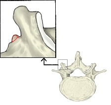 L4
Ala of sacrum (Latin: ala ossis sacri)
The wing-like upper surface of the lateral part of the sacrum on each side of the base. It is formed from transverse processes and rib elements. The figure marks one side, roughly.
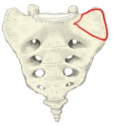 Sacrum (anterior view)
Anterior arch of atlas (Latin: arcus anterior atlantis)
The arched part of the atlas in front of the lateral masses. The extent marked in red is approximate.
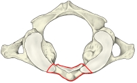 Atlas
Anterior sacral foramina (Latin: foramina sacralia anteriora)
Four pairs of openings on the front of the sacrum, lateral to the transverse ridges; the anterior rami of the sacral nerves leave through them. The figure marks one side.
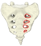 Sacrum (anterior view)
Anterior tubercle of atlas (Latin: tuberculum anterius atlantis)
A small tubercle in the middle of the front of the anterior arch of the atlas; the longus colli attaches to it.
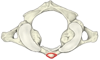 Atlas
Apex of sacrum (Latin: apex ossis sacri)
The lower end of the sacrum, formed by the fifth sacral vertebra. Its lower surface is a transversely oval facet joined to the coccyx, either through an intervertebral disc (sacrococcygeal joint) or by bony fusion.
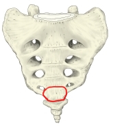 Sacrum (anterior view)
Atlas (Latin: atlas)
The first cervical vertebra. It has no body and no spinous process, so its shape differs greatly from that of other vertebrae.
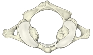 Atlas (superior view)
Auricular surface of sacrum (Latin: facies auricularis ossis sacri)
The ear-shaped surface on each lateral part of the sacrum, which articulates with the auricular surface of the ilium. The joint is the sacroiliac joint; it allows very little movement. The right figure marks one side.
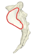 Sacrum (lateral view)
 Sacrum (posterior view)
Axis (Latin: axis)
The second cervical vertebra, characterized by the dens, which projects upward from its body.
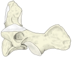 Axis (left lateral view)
Base of sacrum (Latin: basis ossis sacri)
The oval surface at the upper end of the sacrum; it is joined to L5 by an intervertebral disc.
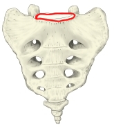 Sacrum (anterior view)
Coccygeal cornu (Latin: cornu coccygeum)
One of the two processes projecting upward from the part of the coccyx that corresponds to the first coccygeal vertebra; they correspond to superior articular processes. The figure marks one side.
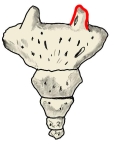 Coccyx (posterior view)
Costal facets (Latin: fovea costalis)
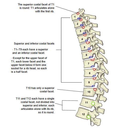
The articular facets on the sides of the bodies of the thoracic vertebrae for the heads of the ribs. Their position and number vary:
· T1–T9: two on each side, a superior and an inferior costal facet.
· T10: only a superior costal facet.
· T11 and T12: a single costal facet each, not divided into superior and inferior.
The superior costal facet of T1 and the facets of T11 and T12 each receive a rib on their own, so they are complete round facets; the others are half facets.
 Costal facets explained
Costal process (Latin: processus costiformis)
A flat process projecting laterally from the side of the pedicle of a lumbar vertebra. It represents the rib of this region and is usually called the transverse process in English texts. Its thickness and length differ from vertebra to vertebra.
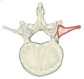 L2
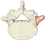 L5
Dens (Latin: dens axis)
The process projecting upward from the body of the axis. It has an anterior and a posterior articular facet and articulates with the atlas (median atlanto-axial joint).
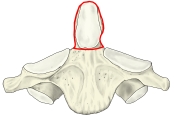 Axis (anterior view)
Facet for dens (Latin: fovea dentis)
A smooth small facet on the back of the anterior arch of the atlas; it articulates with the anterior articular facet of the dens of the axis (median atlanto-axial joint).
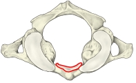 Atlas
Groove for vertebral artery (Latin: sulcus arteriae vertebralis)
A groove on the upper surface of the atlas, where the posterior arch joins the lateral mass. The vertebral artery and the suboccipital nerve run in it. The figure marks one side.
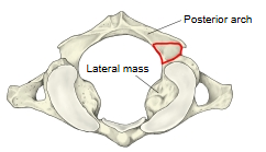 Atlas (superior view)
Inferior articular facet (Latin: facies articularis inferior)
The articular facet on the end of the inferior articular process; it articulates with the superior articular facet of the vertebra below (zygapophysial joint). In the cervical and thoracic vertebrae it faces mainly forward (and downward); from T12 down it faces mainly laterally. For the atlas some texts use the name inferior articular fossa; others call it the inferior articular facet as for other vertebrae.
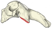 C4
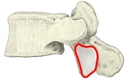 L5
Inferior articular facet of atlas (Latin: facies articularis inferior atlantis)
The articular facet on the lower surface of the lateral mass of the atlas; it articulates with the superior articular facet of the axis (lateral atlanto-axial joint). Some texts call it the inferior articular fossa.
Inferior articular process (Latin: processus articularis inferior)
The process projecting downward from each side of the vertebral arch near its root; it carries the inferior articular facet. How far it projects differs by region: in the cervical and upper thoracic vertebrae it is short, in the lumbar vertebrae it is longer. The extent marked in red is approximate.
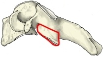 C4
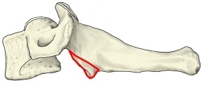 C7
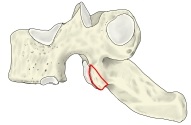 T2
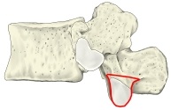 T12
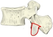 L2
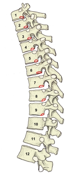
Inferior costal facet (Latin: fovea costalis inferior)
The articular facet at the lower back of each side of the bodies of T1–T9. Together with the superior costal facet of the vertebra below, it articulates with the head of a rib (joint of head of rib).
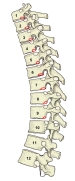 T1–T12
Intermediate sacral crest (Latin: crista sacralis medialis)
A ridge on the back of the sacrum just lateral to the median sacral crest (medial to the posterior sacral foramina), formed by the fused articular processes. Also called the medial sacral crest. The figure marks one side; the upper and lower ends are approximate.
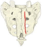 Sacrum (posterior view)
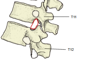
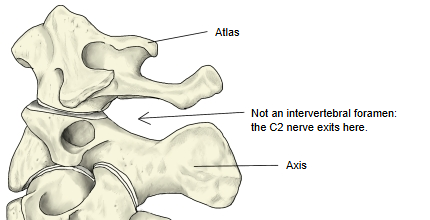
Intervertebral foramen (Latin: foramen intervertebrale)
The opening between two adjacent vertebrae, bounded by the inferior vertebral notch of the vertebra above and the superior vertebral notch of the vertebra below; a spinal nerve leaves through it.
The first and second cervical nerves do not pass through true intervertebral foramina: C1 leaves above the posterior arch of the atlas and C2 between the atlas and the axis. The first true intervertebral foramen lies between C2 and C3.
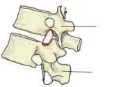 T11–T12
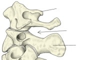 Atlas and axis
Lamina of vertebral arch (Latin: lamina arcus vertebrae)
The arched part of the vertebral arch behind the articular processes. The extent marked in red is approximate.
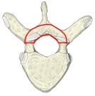 T6
Lateral mass of atlas (Latin: massa lateralis atlantis)
The greatly thickened part of the atlas where the anterior and posterior arches meet. It corresponds to the superior and inferior articular processes of other vertebrae and likewise carries articular facets above and below, the superior and inferior articular facets (fossae). The figure marks one side, roughly.
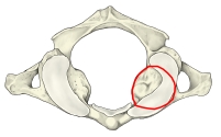 Atlas
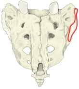
Lateral sacral crest (Latin: crista sacralis lateralis)
The outermost ridge on the back of the sacrum (lateral to the posterior sacral foramina), formed by the fused transverse processes. There is one on each side; the figure marks the right one.
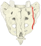 Sacrum (posterior view)
Mammillary process (Latin: processus mammillaris)
A small rounded tubercle on the posterior border of the superior articular process of a lumbar vertebra (also on T12). With the accessory process it is regarded as part of the original transverse process.
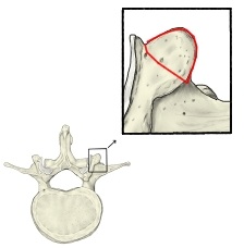 L2
Medial sacral crest (Latin: crista sacralis medialis)
Another name for the intermediate sacral crest. Atlases and textbooks use either name.
- Sacrum (posterior view)
Median sacral crest (Latin: crista sacralis mediana)
The midline ridge on the back of the sacrum, formed by the fused spinous processes of the sacral vertebrae; it shows four tubercles.
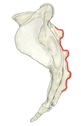 Sacrum (lateral view)
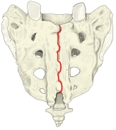 Sacrum (posterior view)
Pedicle of vertebral arch (Latin: pediculus arcus vertebrae)
The short, fairly straight part of the vertebral arch between the body and the articular processes. The figure marks one side, roughly.
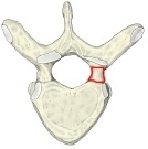 T6
Posterior arch of atlas (Latin: arcus posterior atlantis)
The arched part of the atlas behind the lateral masses. The extent marked in red is approximate.
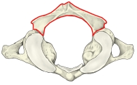 Atlas
Posterior sacral foramina (Latin: foramina sacralia posteriora)
Four pairs of openings just lateral to the intermediate sacral crests; the posterior rami of the sacral nerves pass through them. The figure marks the right side.
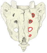 Sacrum (posterior view)
Posterior tubercle of atlas (Latin: tuberculum posterius atlantis)
A small elevation in the middle of the posterior arch of the atlas, in place of a spinous process; the rectus capitis posterior minor arises from it.
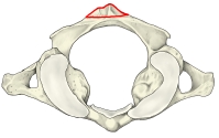 Atlas
Promontory (Latin: promontorium)
The front edge of the base of the sacrum, which projects strongly forward.
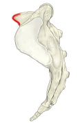 Sacrum (lateral view)
Sacral canal (Latin: canalis sacralis)
The canal running lengthwise through the sacrum, the lower continuation of the vertebral canal. It contains the cauda equina (the roots of the sacral and coccygeal nerves). The lower end shown in the figure is approximate.
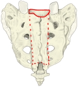 Sacrum (posterior view)
Sacral cornu (Latin: cornu sacrale)
The lower end of each intermediate sacral crest, projecting downward like a horn. The figure marks the right one; the extent may be inexact.
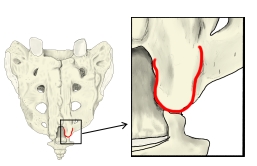 Sacrum (posterior view, inset enlarged)
Sacral hiatus (Latin: hiatus sacralis)
The opening at the lower end of the sacral canal on the back of the sacrum, where the laminae of the fifth (and sometimes fourth) sacral vertebra have not fused. Its apex usually lies at the level of the fourth sacral vertebra.
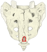 Sacrum (posterior view)
Sacral tuberosity (Latin: tuberositas ossis sacri)
The rough area on the back of the lateral part of the sacrum; the interosseous sacroiliac ligament attaches to it. The figure marks the right side, roughly.
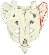 Sacrum (posterior view)
Spinous process (Latin: processus spinosus)
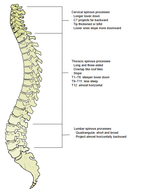
The process projecting backward from the vertebral arch of every vertebra except the atlas. How far and in which direction it projects differs by region; some have a bifid tip. In the atlas the posterior tubercle takes its place.
[Cervical vertebrae] Longer from above downward. The spinous process of C7 projects far backward and is easily felt under the skin. The tip is thickened into a tubercle or bifid; the lower ones slope more steeply downward.
[Thoracic vertebrae] Long and three-sided, overlapping like roof tiles. T1–T8: steeper lower down; T9–T11: less steep; T12: almost horizontal.
[Lumbar vertebrae] Quadrangular, short and broad; projects almost horizontally backward.
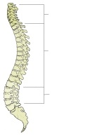 Spinous processes explained
Superior articular facet (Latin: facies articularis superior)
The articular facet on the end of the superior articular process; it articulates with the inferior articular facet of the vertebra above (zygapophysial joint). In the cervical vertebrae it faces upward and backward; in the thoracic vertebrae mainly backward (and laterally); in the lumbar vertebrae mainly medially. For the atlas some texts use the name superior articular fossa.
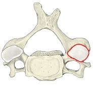 C4
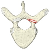 T6
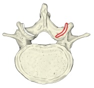 L4
Superior articular facet of atlas (Latin: facies articularis superior atlantis)
The articular facet on the upper surface of the lateral mass of the atlas; it articulates with the occipital condyle of the occipital bone (atlanto-occipital joint). Some texts call it the superior articular fossa. The figure marks one side.
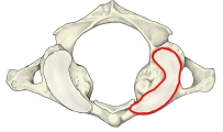 Atlas
Superior articular process (Latin: processus articularis superior)
The process projecting upward from each side of the vertebral arch near its root; it carries the superior articular facet. The extent marked in red is approximate.
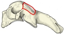 C4
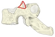 T2
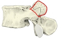 L4
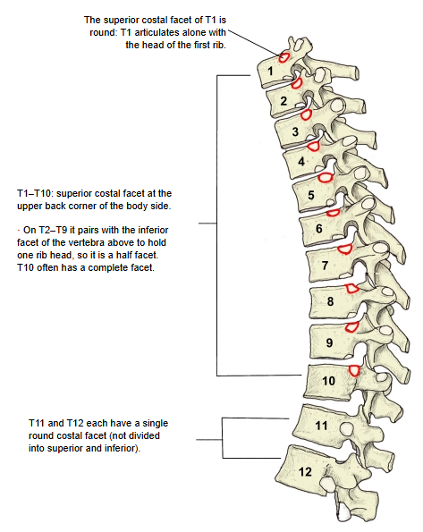
Superior costal facet (Latin: fovea costalis superior)
The articular facet at the upper back of each side of the bodies of T1–T10, on the upper epiphyseal rim. On T2–T9 it joins the inferior costal facet of the vertebra above to receive the head of one rib, so each is a half facet. The superior costal facet of T1 receives the first rib on its own and is a complete round facet; on T10 it is usually also a complete facet.
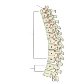 Superior costal facets explained
Transverse costal facet (Latin: fovea costalis processus transversi)
The small articular facet on the tip of the transverse process of T1–T10; it articulates with the tubercle of the rib (costotransverse joint). The 11th and 12th ribs are short and do not reach the transverse processes, so T11 and T12 have no transverse costal facet.
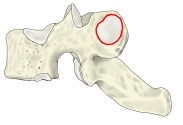 T2
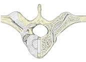 T8 with ribs (transverse section)
Transverse process (Latin: processus transversus)
The process projecting laterally on each side of the vertebral arch, a little in front of the spinous process. Many muscles of the back attach to it. Its shape, size and direction differ by region.
· Cervical vertebrae: it has a transverse foramen. On C3–C7 it ends in an anterior and a posterior tubercle.
· Thoracic vertebrae: it projects backward and laterally; its tip carries the transverse costal facet for the rib, which is absent on T11 and T12.
· Lumbar vertebrae: what projects laterally is the costal process (usually called the transverse process in English texts); the original transverse process is represented by the accessory and mammillary processes.
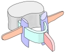 Parts of a vertebra
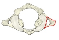 Atlas
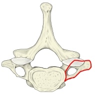 C7
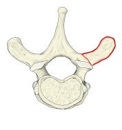 T2
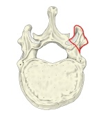 T12
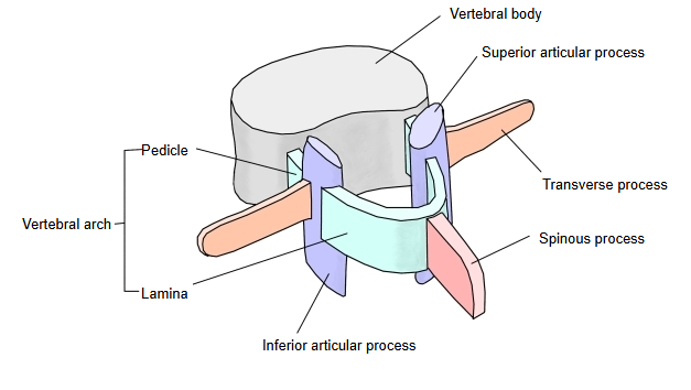
Only one transverse process is marked in red, and its extent is approximate.
Transverse ridges (Latin: lineae transversae)
Four ridges across the middle of the front of the sacrum, marking the boundaries between the fused sacral vertebrae.
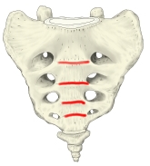 Sacrum (anterior view)
Uncinate process (uncus of body) (Latin: uncus corporis vertebrae)
A raised lip on each side of the upper surface of the bodies of C3–C7 (sometimes T1), where the pedicles meet the body. It articulates with the vertebra above at the small uncovertebral joints. It is found only in the cervical region. The figures mark one side.
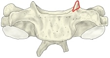 C4 (anterior view)
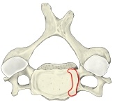 C4 (superior view)
Vertebra prominens (Latin: vertebra prominens)
The seventh cervical vertebra. Its spinous process is especially long and projects far backward, so it is easily felt under the skin.
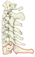 Cervical vertebrae (left lateral view)
Vertebral arch (Latin: arcus vertebrae)
The arched part behind the vertebral body. It is divided into the pedicles and the laminae.
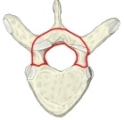 T6
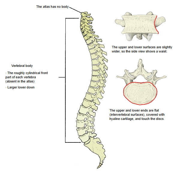
Vertebral body (Latin: corpus vertebrae)
The roughly cylindrical front part of a vertebra.
1. The atlas has no body.
2. The bodies become larger from above downward.
3. The upper and lower surfaces are slightly wider than the middle, so the side view shows a slight waist.
4. The upper and lower ends are flat (intervertebral surfaces), covered with hyaline cartilage, and touch the intervertebral discs. The rim of each surface ossifies as a ring (ring apophysis) that is incomplete at the back; with age, the edges of the bodies grow outward horizontally.
 Vertebral bodies explained
Vertebral foramen (Latin: foramen vertebrale)
The opening between the vertebral arch and the vertebral body; in the atlas, which has no body, it is enclosed by the anterior and posterior arches. The foramina together form the vertebral canal, which contains the spinal cord. Its shape differs by region.
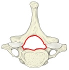 C7
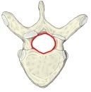 T6
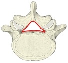 L5
Vertebral column: Overview | Features of the vertebrae | Related terms | Ligaments and membranes | Joints
References and sources
- FIPAT. Terminologia Anatomica, 2nd ed. (TA2), 2019.
- Gray H. Anatomy of the Human Body, 20th ed. (1918): The vertebral column.