Liver (Latin: hepar)
- Overview
- Location
- Surfaces (diaphragmatic surface, visceral surface)
- Divisions (anatomical division, functional division: Couinaud classification, Healey–Schroy classification)
- Structure
- Functions
- Vessels and nerves (arteries / veins / lymphatics / nerves / bile ducts)
- Related terms
- References
Overview
· Weight: 1,000–1,300 g in men, 900–1,000 g in women.
The glands belonging to the digestive system are shown briefly below.
the digestive
system
- Salivary glands : around the oral cavity; divided into the major salivary glands (parotid gland and others) and the minor salivary glands (labial glands, lingual glands and others).
- Gastric glands : secrete gastric juice (gastric acid, pepsin) from the mucosa; work: digesting proteins, killing germs, aiding digestion.
- Liver : produces bile (aiding the digestion of fats); stored and concentrated in the gallbladder and secreted into the duodenum.
- Pancreas : as an exocrine gland it secretes mainly digestive enzymes; as an endocrine gland it secretes hormones.
- Duodenal glands : = Brunner's glands; in the submucosa of the duodenum; secrete alkaline fluid rich in mucus.
- Intestinal glands : in the walls of the small and large intestines; secrete digestive enzymes (peptidases, maltase) and mucus.
Location
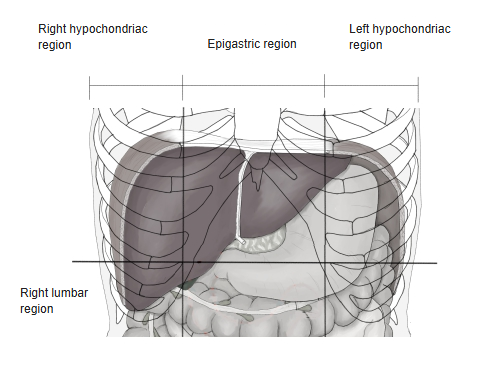
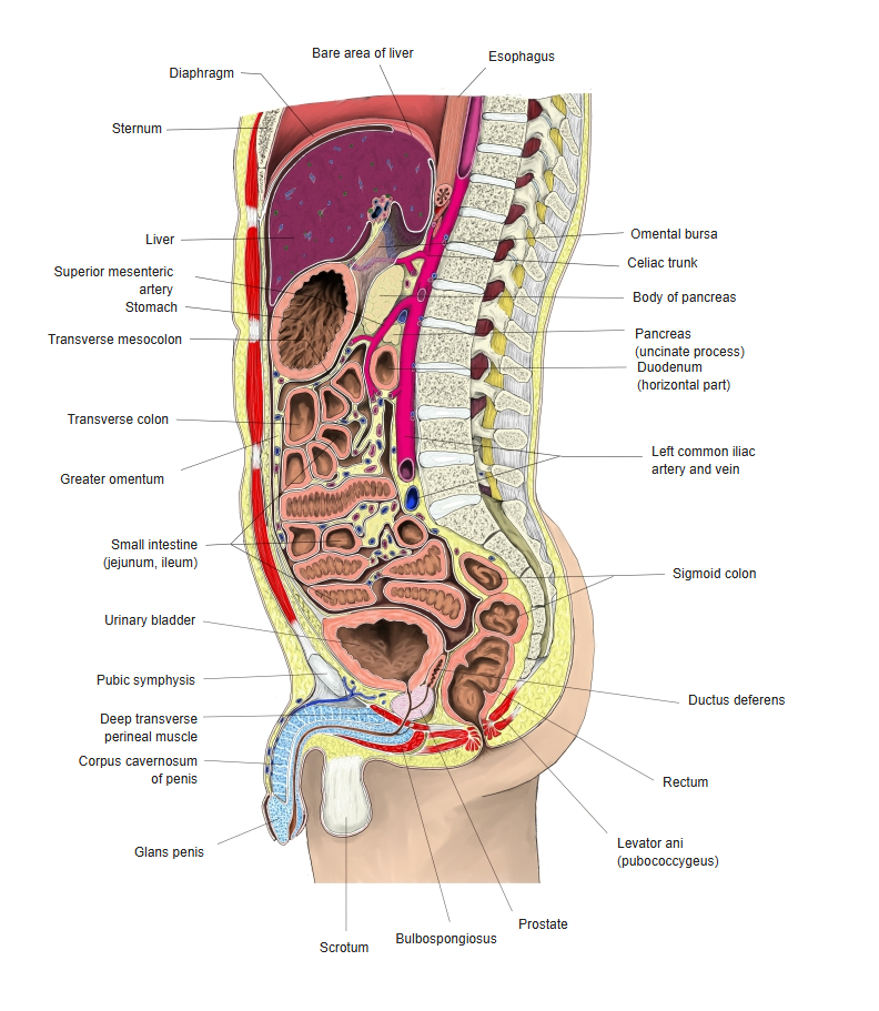
· The liver lies in the upper abdomen and both hypochondriac regions, attached to the domes of the diaphragm; its middle part touches the central tendon of the diaphragm, and its back touches the lumbar part of the diaphragm.
· Height: it changes with posture and breathing, moving about 2–3 cm even in quiet breathing.
· The back of the liver lies at the level of the 9th–10th thoracic vertebrae; at expiration its highest point reaches the 5th intercostal space on the right.
· The lower border of the liver runs along the right costal arch in front, crosses obliquely from lower right to upper left between the two costal arches to reach the left 7th costal cartilage, and in the midline lies about three finger-breadths below the xiphoid process.
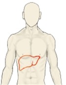 Position seen from the body surface
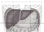 Abdominal cavity and liver
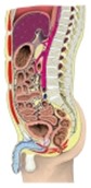 Trunk (sagittal section)
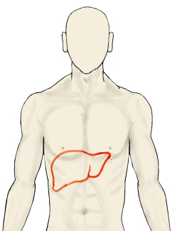
Surfaces
of the liver
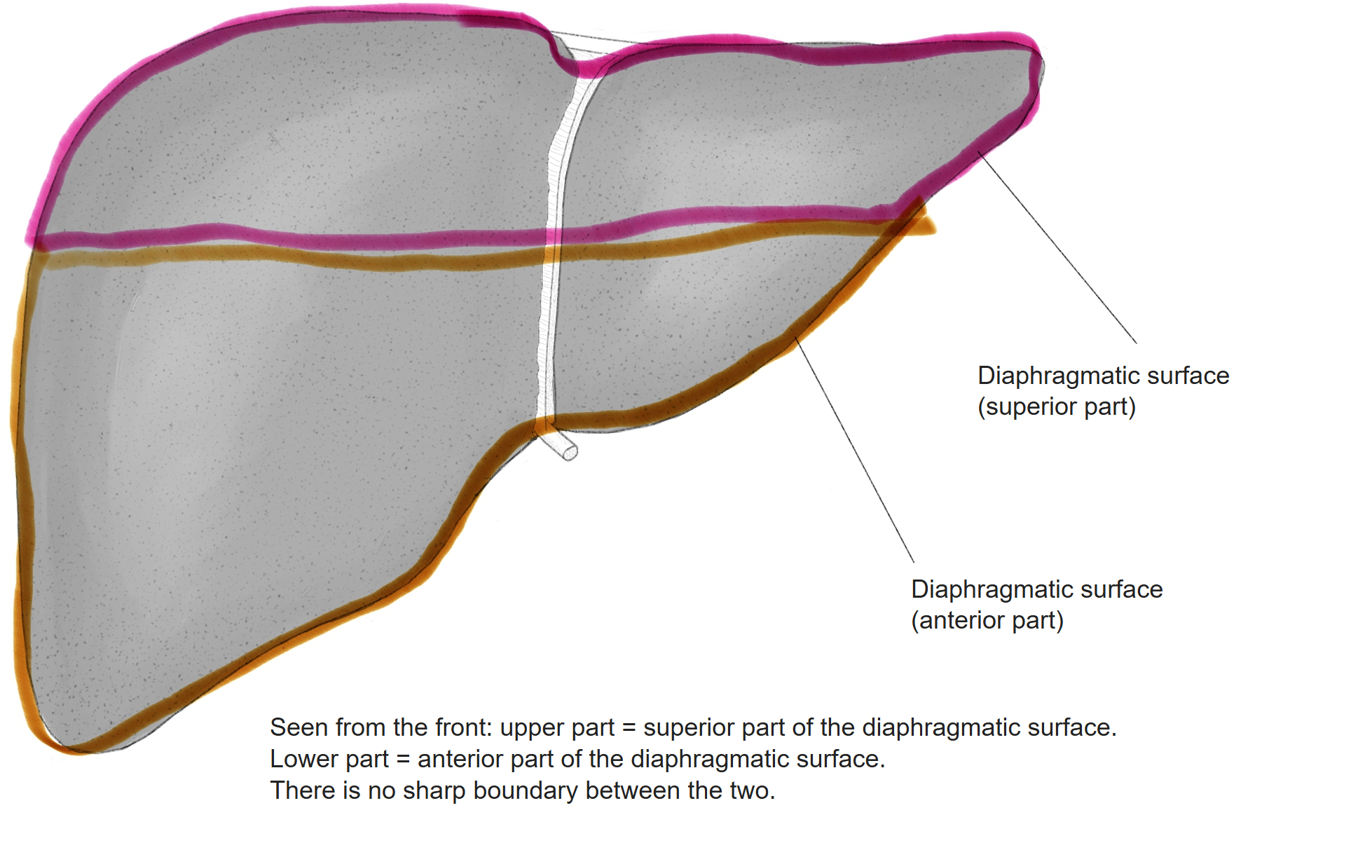
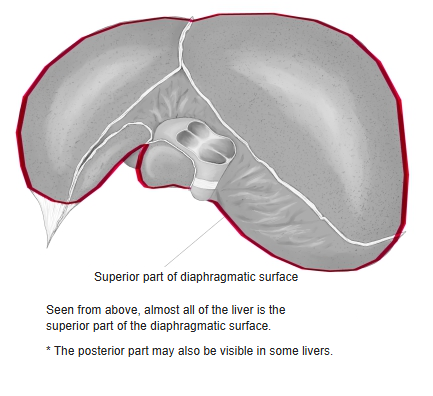
- Diaphragmatic surface = upper surface
- Posterior part : mostly the bare area (covered by fibrous membrane, without peritoneum), fused to the underside of the diaphragm.
- Superior part : convex (especially on the right); the falciform ligament is seen in front.
- Anterior part : the falciform ligament continuing from the upper surface runs down it and divides the liver into 2 lobes.
- Visceral surface = lower surface : covered by peritoneum except for some parts (porta hepatis, fissure for the ligamentum teres, gallbladder fossa). Concave on the whole, but with local hollows (impressions). Vessels, nerves, bile ducts and lymph vessels enter and leave (porta hepatis).
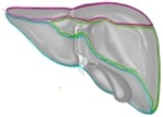
The surfaces above are colour-coded in the illustrations below; few sources were available for reference, so they may not be fully accurate.
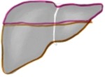 Liver from the front
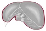 Liver from above
 Liver from behind
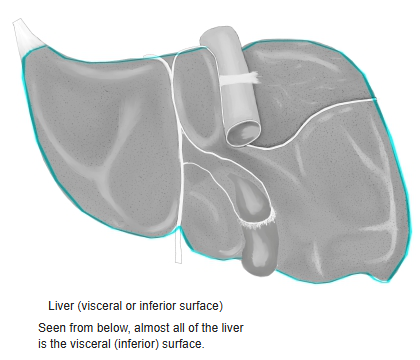
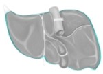 Liver from below
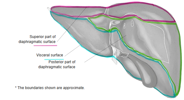
■ Diaphragmatic surface (upper surface) ■
[Posterior part]
· It is fused to the underside of the diaphragm, and most of it lacks peritoneum.
· It is roughly triangular and is also called the bare area.
· The bare area is roughly triangular and is bounded by the coronary ligament and the triangular ligaments.
[Superior part]
· A convex surface touching the diaphragm, especially marked on the right half.
· The falciform ligament, a fold in line with the midline of the body, is seen and divides the liver into 2 lobes.
· The shallow hollow made by the heart, to the left of and in front of the inferior vena cava, is called the cardiac impression.
[Anterior part]
· Covered by peritoneum except where the falciform ligament is attached.
· The falciform ligament, continuing from the upper surface, runs down and divides the liver into the right lobe and left lobe.
· Most of it touches the diaphragm.
· Through the diaphragm it lies at the level of the pleura (costodiaphragmatic recess) and the 6th–10th ribs on the right, and of the 7th–8th costal cartilages on the left.
· The middle of the anterior part lies behind the xiphoid process.
· The right anterior part lies at the level of the 7th–11th ribs and touches the right lung and pleura through the diaphragm.
■ Visceral surface (lower surface) ■
· Covered by peritoneum except for some parts (porta hepatis, fissure for the ligamentum teres, gallbladder fossa).
· Concave on the whole, but with local hollows (impressions) where other organs touch it: gastric, duodenal, colic, renal and suprarenal impressions.
· The middle part is deeply hollowed from side to side, and the portal vein, proper hepatic artery, hepatic ducts, nerves and lymph vessels enter and leave it (porta hepatis). The porta hepatis is joined to the upper part of the duodenum by the hepatoduodenal ligament, in which the portal vein, proper hepatic artery and common bile duct run.
· Two grooves (left and right sagittal fossae) run sagittally on either side of the porta hepatis, forming an H with it.
· The front of the right sagittal fossa is the gallbladder fossa, holding the gallbladder; its back is the groove for the vena cava, holding the inferior vena cava.
· The left sagittal fossa runs without a break; in front is the fissure for the ligamentum teres (continuing from the notch for the ligamentum teres), and behind is the fissure for the ligamentum venosum.
 Liver, visceral surface 1
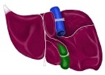 Liver, visceral surface 2
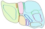 Liver, visceral surface (schematic)
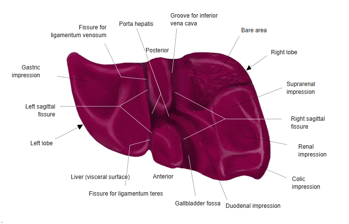
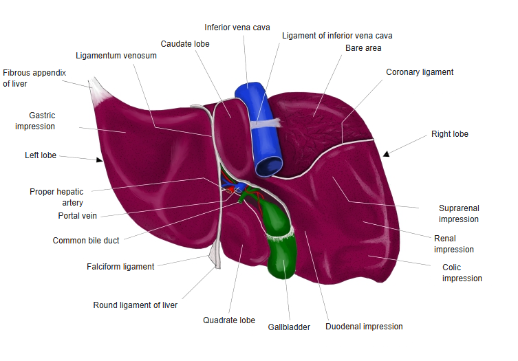
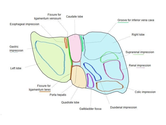
on the visceral
surface
- Right lobe
- Suprarenal impression : the hollow made by the right suprarenal gland; partly or wholly in the bare area.
- Renal impression : the hollow made by the right kidney.
- Colic impression : the hollow made by the colon, on the right of the gallbladder.
- Duodenal impression : the hollow made by the duodenum, on the right near the neck of the gallbladder.
- Left lobe
- Esophageal impression : the hollow made by the esophagus, but apparently not always present.
- Gastric impression : the hollow made by the stomach, the largest of the impressions.
Divisions
There are 2 main ways of dividing the liver: anatomically into left and right lobes, and more finely, based mainly on the function of the vessels.
■ Anatomical division ■
· This division is said to be unrelated to the functions of the liver itself.
The right lobe is further divided into 3 parts as follows.
right lobe
- Right lobe (in the narrow sense) : the part excluding the quadrate and caudate lobes below.
- Quadrate lobe : the four-sided part at the front of the lower surface, surrounded by the fissure for the ligamentum teres, the gallbladder fossa and the porta hepatis.
- Caudate lobe : the part at the back, surrounded by the groove for the vena cava, the fissure for the ligamentum venosum and the porta hepatis.
Border: on the front, the falciform ligament running down; on the lower surface, the left sagittal fossa forms the border between the 2 lobes.
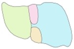 Right and left lobes (colour-coded)
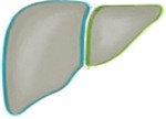 Liver (front)
- Parts of the visceral surface
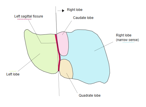
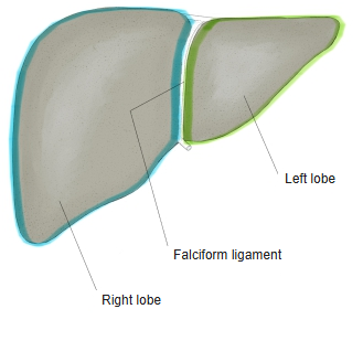
■ Functional division ■
· The ratio of the functional right lobe to the left lobe is about 2:1.
the two lobes
- Anatomical division ⇒ falciform ligament : a line formed where 2 layers of peritoneum meet, running down the front of the liver.
- Functional division ⇒ Cantlie's line : an imaginary line on the diaphragmatic surface joining the gallbladder fossa and the inferior vena cava.
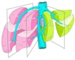 Portal fissures
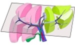 Horizontal portal plane?
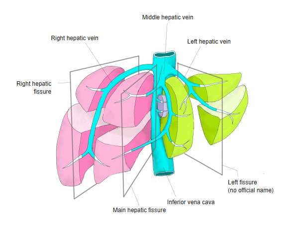
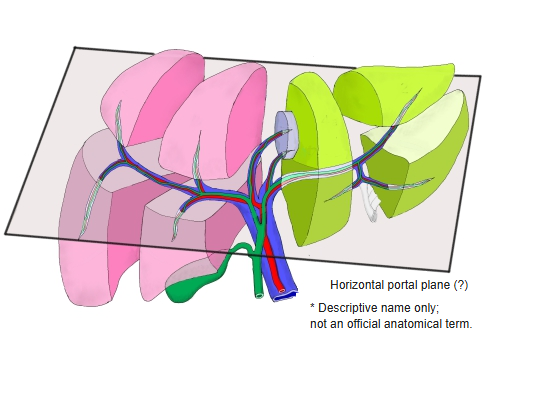
[Division into 3 by the fissures]
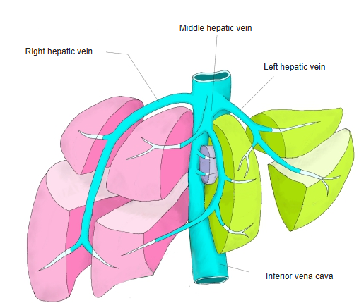
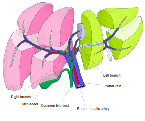
· The liver is divided vertically into 4 by the right, middle and left hepatic veins, and these parts are further divided according to the branches of the portal vein, proper hepatic artery and bile ducts running from side to side.
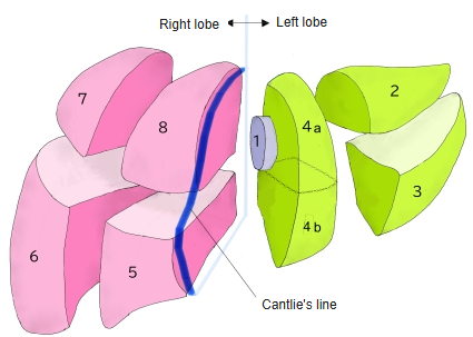
· The border between the right and left lobes is Cantlie's line (joining the gallbladder fossa and the inferior vena cava on the diaphragmatic surface), which differs from the anatomical division (falciform ligament).
· The caudate lobe is regarded as an independent part belonging to neither lobe, and is sometimes divided into 3 (Healey–Schroy classification).
[Couinaud classification]
segments
- Right lobe
- Anterior sector : right anterior inferior segment (V), right anterior superior segment (VIII) → lie in the lower and upper parts of the liver.
- Posterior sector : right posterior inferior segment (VI), right posterior superior segment (VII).
- Caudate lobe (I) : regarded as an independent lobe belonging to neither the right nor the left lobe.
- Left lobe
- Medial sector : left medial segment (IV) : to the right of the falciform ligament; sometimes divided into upper and lower parts.
- Lateral sector : left lateral anterior segment (III), left lateral posterior segment (II) : to the left of the falciform ligament; upper part → posterior segment, lower part → anterior segment.
- The border between the right and left lobes is Cantlie's line.
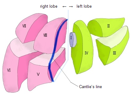
 Hepatic segments and Cantlie's line
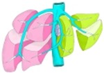 Hepatic segments and hepatic veins
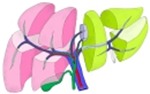 Hepatic segments and portal vein
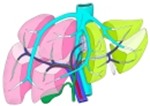 Hepatic segments and vessels
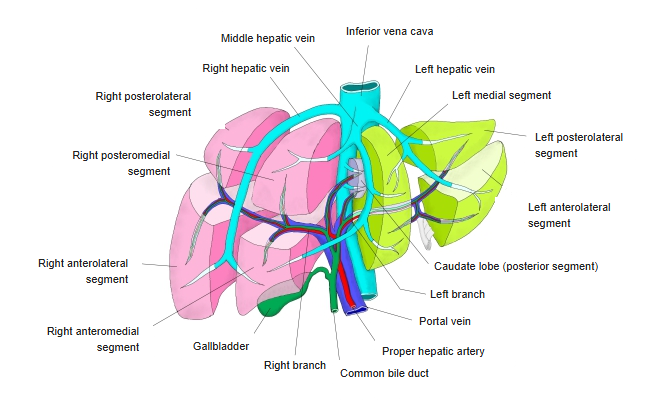
[Healey–Schroy classification]
segments
- Right lobe
- Anterior segment : superior area, inferior area
- Posterior segment : superior area, inferior area
- Caudate lobe : left part, right part, caudate process
- Left lobe
- Medial segment : superior area, inferior area
- Lateral segment : superior area, inferior area
- The border between the right and left lobes is Cantlie's line.
Note: the Healey–Schroy system names the parts by position (no Roman numerals); its divisions roughly correspond to Couinaud's segments.
Structure
of the liver
- Parenchyma : Hepatocytes
- 70–80% of the liver; about 250–300 billion in all.
- Shape: polygonal; 30–40% have 2 nuclei.
- Average lifespan about 6–10 months.
- Arranged in the hepatic lobules as rows called hepatic plates.
- Work: metabolism of nutrients, detoxification, synthesis of proteins, production of bile.
- Interstitium
- Connective tissue : Glisson's sheath; wraps the vessels and bile ducts running inside the liver.
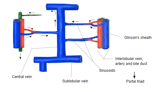
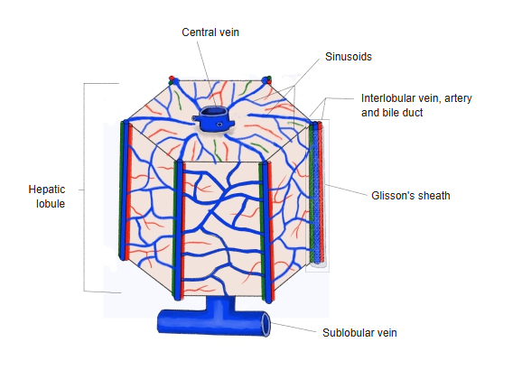
- Vessels : portal vein and proper hepatic artery; finally they join within the hepatic lobule and leave to the inferior vena cava through the hepatic veins.
- Bile ducts : collect the bile made by the hepatocytes and carry it out of the liver; bile canaliculi, interlobular bile ducts and so on.
- Lymph vessels : the liver is the largest producer of lymph; lymph in the hepatic lobules gathers in the lymph vessels in Glisson's sheath.
- Nerves : sympathetic (from the celiac plexus), parasympathetic (vagus nerve).
- Non-parenchymal cells : Kupffer cells, hepatic stellate cells, sinusoidal endothelial cells, bile duct epithelial cells and others.
and its
membranes
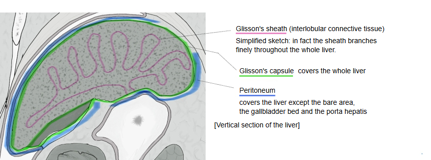
- Surface
- Glisson's capsule : the connective-tissue membrane covering the whole surface; a few cells and nerves are present.
- Peritoneum : covers the surface except for some parts (porta hepatis, bare area, where the gallbladder touches). A serous membrane.
- Parenchyma
- Glisson's sheath : continuous with Glisson's capsule; enters at the porta hepatis and wraps the arteries, veins and bile ducts together.
 The liver and its membranes
Functions
The work of the hepatocytes is briefly as follows.
1. Metabolism: processes, stores and converts the nutrients absorbed from food (sugars, fats, proteins). Conversion: changes glucose into glycogen for storage.
2. Detoxification: breaks down and renders harmless substances harmful to the body (alcohol, drugs, ammonia and so on).
3. Production of bile: makes bile from cholesterol, bilirubin (a breakdown product of haemoglobin) and other substances, and sends it to the gallbladder.
4. Synthesis of blood components: makes albumin (a blood protein) and blood-clotting factors.
[Other cells]
The main cells of the liver are the hepatocytes, but the following are also components.
| Name | Description | |
|---|---|---|
| 1 | Kupffer cells | Immune cells of the liver; they engulf foreign matter, bacteria and old red blood cells. Number: about 15% of the liver cells. |
| 2 | Hepatic stellate cells | Lie between the sinusoids and the hepatocytes (space of Disse); store vitamin A and so on. |
| 3 | Sinusoidal endothelial cells | Form the sinusoids; unlike ordinary endothelial cells they are not continuous and have many small pores (fenestrations). |
| 4 | Bile duct epithelial cells | Form the walls of the bile ducts. |
| 5 | Others | Immune cells (T cells, NK cells and others), liver progenitor cells, fibroblast-like cells, vascular endothelial cells (portal vein, hepatic artery and others). |
Vessels and nerves
[Arteries]
Proper hepatic artery: one of the branches of the common hepatic artery; at the porta hepatis it divides into right and left branches, which enter the liver.
hepatic
artery
- Gastroduodenal artery
- Proper hepatic artery
- Right branch (to the right lobe) : cystic artery (also supplies the lower surface of the liver and the cystic duct), anterior segmental artery, posterior segmental artery, caudate lobe artery.
- Left branch (to the left lobe) : caudate lobe artery, medial segmental artery, lateral segmental artery.
- Right gastric artery : supplies the lesser curvature and pylorus. Note: it sometimes arises from the common hepatic artery and so on.
[Veins]
The main flow of the veins into and out of the liver is as follows.
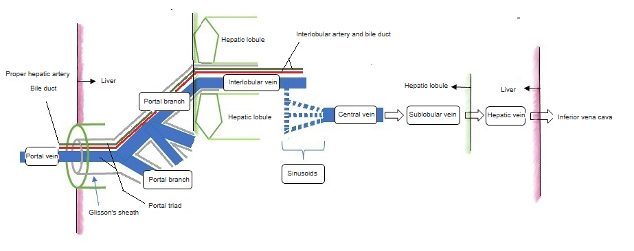
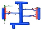 From the interlobular vein to the central vein
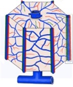 Hepatic lobule and vessels
[Lymphatics]
The lymph vessels (lymphatic system) entering and leaving the liver are one of its important drainage routes; the liver is also the organ that produces the most lymph in the body (said to be about 20–50% of all lymph).
· Superficial lymph vessels · Deep lymph vessels
[Nerves]
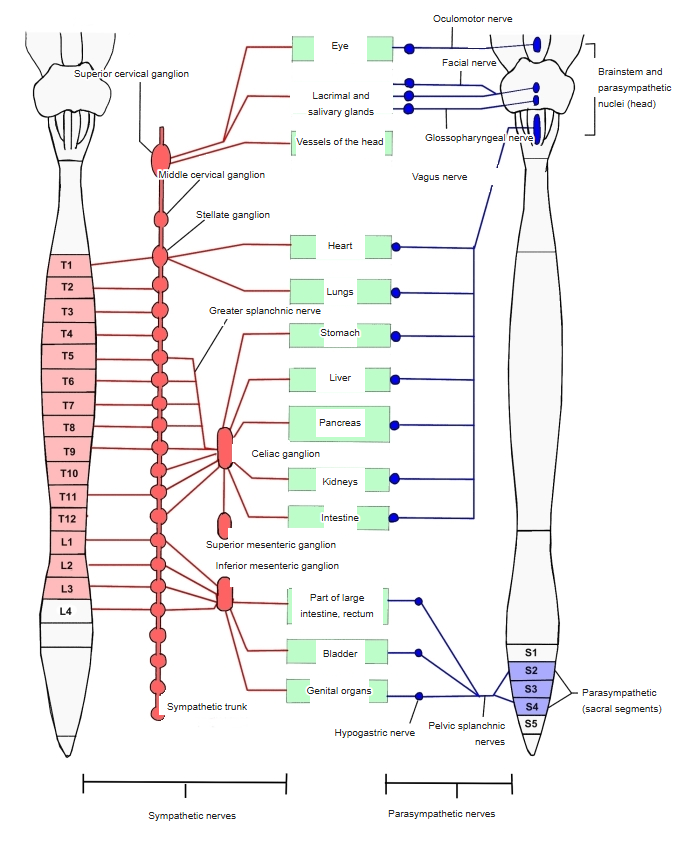
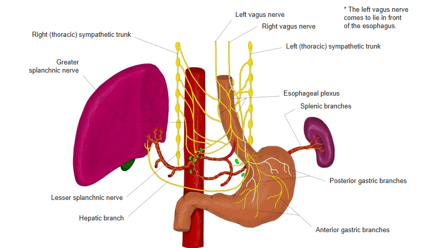
· Sympathetic: nerve fibres from the celiac plexus.
· Parasympathetic: the vagus nerve; fibres from the anterior and posterior vagal trunks form the hepatic plexus as hepatic branches going to the liver.
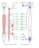 Distribution of the autonomic nervous system
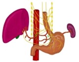 Vagus nerve (thorax and abdomen)
[Hepatic ducts]
The right hepatic duct collects bile from the right lobe of the liver and the left hepatic duct from the left lobe; they leave the parenchyma and join to form the common hepatic duct, which leaves at the porta hepatis.
0.5–1.5 µm⇒Bile ductules
0.5–2 µm⇒Canals of Hering
10–15 µm⇒Interlobular bile ducts
10–30 µm⇒Segmental bile duct branches
1–2 mm⇒Right / left hepatic ducts
4–6 mm⇒out of the liver
· Some sources do not distinguish the first 3 ducts (bile canaliculi, ductules, canals of Hering), and some give the ductules and the canals of Hering in the reverse order.
· The diameters are only a rough guide.
- Intrahepatic bile ducts
- Within the hepatic lobule : Bile canaliculi the finest bile passages, without a special wall of their own; Bile ductules made of flattened bile duct epithelial cells; Canals of Hering link the bile passages in the lobule with the interlobular bile ducts (the order of the last two is reversed in some sources).
- Interlobular bile ducts : receive bile from the hepatic lobules; one of the "portal triad".
- Segmental bile duct branches : gather the bile from the interlobular bile ducts segment by segment.
- Left / right hepatic ducts : gather the bile from the segmental branches for the right and left lobes and leave the liver.
- Extrahepatic bile ducts
- Common hepatic duct : formed by the union of the left and right hepatic ducts.
- Cystic duct : carries bile from the common hepatic duct to the gallbladder (and back).
- Common bile duct : finally carries the bile made in the liver to the duodenum.
Related terms
Main anatomical terms related to the liver, in alphabetical order.
| B | Bare area of liver |
| C | Cardiac impression, Caudate lobe, Caudate process, Central vein, Colic impression, Common bile duct |
| D | Diaphragmatic surface, Duodenal impression |
| E | Esophageal impression |
| F | Fibrous appendix of liver, Fibrous capsule, Fissure for ligamentum teres, Fissure for ligamentum venosum |
| G | Gallbladder fossa, Gastric impression, Glisson's capsule, Glisson's sheath, Groove for vena cava |
| H | Hepatic lobule, Hepatic plates, Hepatic segments |
| I | Interlobular artery, Interlobular bile ducts, Interlobular vein |
| L | Left hepatic duct, Left lobe of liver, Ligamentum venosum, Lobes of liver |
| N | Notch for ligamentum teres |
| O | Omental tuberosity |
| P | Papillary process, Porta hepatis, Portal fissures |
| Q | Quadrate lobe |
| R | Renal impression, Right hepatic duct, Right lobe of liver, Round ligament of liver |
| S | Suprarenal impression |
| U | Umbilical fissure |
| V | Visceral surface |
References and sources
- Further reading: Liver, Wikipedia.