Petrous part (Latin: pars petrosa ossis temporalis)
Also called the petrous part of the temporal bone or petrous bone
- Squamous part : occupies the middle and the whole upper part of the bone; it has two surfaces, temporal and cerebral.
- Petrous part
- Mastoid part : below the squamous part, from the mastoid process up to the parietal notch.Related terms: mastoid process, mastoid notch, groove for occipital artery, groove for sigmoid sinus, mastoid foramen, occipital border, stylomastoid foramen, canaliculus for chorda tympani, petrotympanic fissure
- Pyramid : a three-sided pyramid running forwards and medially from the boundary of the squamous and mastoid parts, with three surfaces and three borders: the petrous part in the narrow sense.Related terms: apex, foramen lacerum, sphenopetrosal fissure, arcuate eminence, tegmen tympani, trigeminal impression, groove and hiatus for greater petrosal nerve, groove and hiatus for lesser petrosal nerve, groove for superior petrosal sinus, internal acoustic opening, petro-occipital fissure, groove for inferior petrosal sinus, jugular notch, intrajugular process, petrosquamous fissure, musculotubal canal, septum of musculotubal canal, canal for tensor tympani, canal for auditory tube, jugular fossa, carotid canal, petrous fossula, tympanic canaliculus, cochlear canaliculus, mastoid canaliculus, styloid process, stylomastoid foramen, petrotympanic fissure, tympanosquamous fissure
- Tympanic part : the smallest part: a thin, half-tube-shaped plate that forms the front and lower walls of the external acoustic meatus.
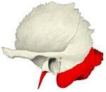 Temporal bone (lateral view)
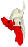 Temporal bone (inferior view)
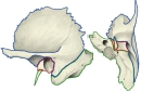 Parts of the temporal bone
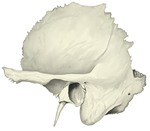 Temporal bone (lateral view), labelled
 Temporal bone (inferior view), labelled
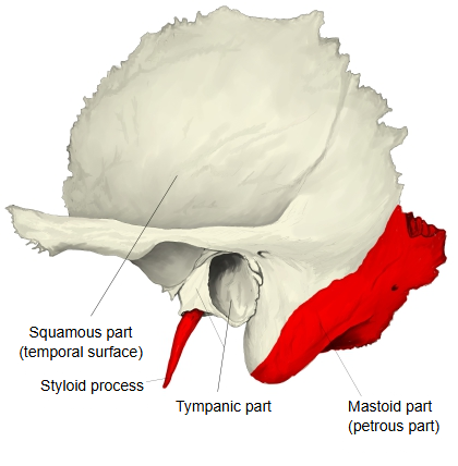
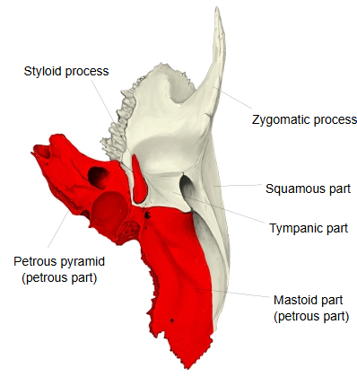
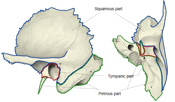
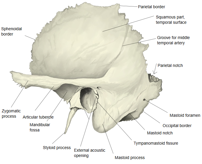
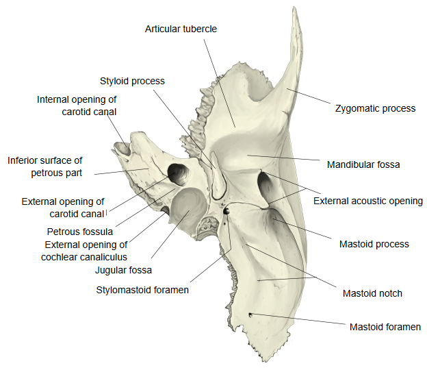
In the wider sense the petrous part (also called the petromastoid part) includes the mastoid part, which projects downwards behind the external acoustic opening as the mastoid process and reaches up to the parietal notch, and the pyramid, a three-sided pyramid of bone that projects forwards and medially from the mastoid part; the pyramid alone is sometimes called the petrous part in the narrow sense. Some texts use petrous part for the pyramid only; sources differ. The whole develops as one piece of bone from the ear capsule of the cartilaginous cranial base, but the two parts are described separately here.
Mastoid part: its outer surface is rough for muscle attachments. The part that extends downwards behind the external acoustic opening is the mastoid process, into which the sternocleidomastoid is inserted (also splenius capitis and longissimus capitis). On the medial side of the process is the mastoid notch, from which the posterior belly of the digastric arises, and medial to it the groove for the occipital artery. On the inner surface runs the deep, long groove for the sigmoid sinus, which continues above into the groove for the transverse sinus and below to the jugular foramen. The mastoid foramen near the back border carries the mastoid emissary vein into this groove. The back border, which joins the occipital squama, is called the occipital border. Inside, the mastoid part contains the mastoid air cells, which drain into the mastoid antrum and so into the tympanic cavity.
Facial canal: the facial canal, the passage of the facial nerve, enters the bone at the facial nerve area of the fundus of the internal acoustic meatus and runs almost horizontally forwards and laterally beside the cochlea. It then turns sharply backwards and laterally, forming the geniculum (knee) of the facial canal, runs back above the oval window between the tympanic cavity and the semicircular canals, and finally curves downwards to open at the stylomastoid foramen. A little above this foramen the canaliculus for the chorda tympani leaves the facial canal; the chorda tympani crosses the tympanic cavity between the handle of the malleus and the long limb of the incus and leaves the skull through the petrotympanic fissure. Fractures of the petrous part can injure the facial nerve.
Pyramid: a three-sided pyramid set obliquely between the sphenoid and occipital bones, running from back and lateral to front and medial; it is one of the densest bones of the body (Latin "petrosus", stony) and houses the inner ear (cochlea, vestibule and semicircular canals) and most of the tympanic cavity. It has three surfaces (anterior, posterior, inferior) and three borders (superior, anterior, posterior). Its tip, the apex, forms the foramen lacerum with the body and greater wing of the sphenoid and the basilar part of the occipital bone; the foramen is closed by fibrocartilage; the greater and deep petrosal nerves do not pass down through it but cross it, joining to form the nerve of the pterygoid canal, which runs forwards into the pterygoid canal. The internal opening of the carotid canal lies at the apex; its external opening is on the under side, and from it two caroticotympanic canaliculi lead into the tympanic cavity. The anterior border forms the sphenopetrosal fissure with the greater wing of the sphenoid.
The anterior surface (cerebral surface) carries the arcuate eminence, the tegmen tympani between the eminence and the petrosquamous fissure, the trigeminal impression near the apex and the grooves for the greater and lesser petrosal nerves. The posterior surface (cerebellar surface) carries the internal acoustic opening, the subarcuate fossa and the external opening of the vestibular aqueduct. On the inferior surface lie the musculotubal canal, the jugular fossa, the external opening of the carotid canal, the petrous fossula, the cochlear canaliculus, the mastoid canaliculus, the styloid process and the stylomastoid foramen. In the mandibular fossa the edge of the tegmen tympani divides the medial part of the tympanosquamous fissure into the petrosquamous fissure in front and the petrotympanic fissure behind. These features are described in more detail below.
Reference websites
[Wikipedia]
· Petrous part of the temporal bone, Wikipedia.
· Mastoid part of the temporal bone, Wikipedia.
[Websites with illustrations]
· The temporal bone (TeachMeAnatomy)
· Temporal bone: anatomy, parts, sutures and foramina (Kenhub)
· Left temporal bone, inner surface (Gray's Anatomy, fig. 138) (Wikimedia Commons)

· Left temporal bone, inferior surface (Gray's Anatomy, fig. 141) (Wikimedia Commons)

Base
The base of the pyramid is fused with the inner surfaces of the squamous part and the mastoid part.
Apex
The apex is rough and uneven. It fits into the angle between the back border of the greater wing of the sphenoid bone and the basilar part of the occipital bone. It carries the internal opening of the carotid canal and forms the back and outer edge of the foramen lacerum.
Surfaces
Anterior surface: it forms the back part of the floor of the middle cranial fossa and is continuous with the cerebral surface of the squamous part at the petrosquamous fissure, which can still be seen late in life. It carries impressions of the brain and the following features:
near its centre, the arcuate eminence, a rounded bulge over the anterior (superior) semicircular canal;
in front of and lateral to the eminence, the tegmen tympani, a very thin plate of bone that forms the roof of the tympanic cavity and separates it from the cranial cavity;
a shallow groove, the groove for the greater petrosal nerve, leading backwards and laterally to the hiatus for the greater petrosal nerve, which carries the greater petrosal nerve and the petrosal branch of the middle meningeal artery;
lateral to it, a smaller groove and hiatus for the lesser petrosal nerve, for the lesser petrosal nerve;
near the apex, the end of the carotid canal, whose wall is incomplete in front;
above it, the shallow trigeminal impression for the trigeminal ganglion.
Posterior surface: it faces the posterior cranial fossa. Near its middle is the internal acoustic opening, which leads into the internal acoustic meatus for the facial and vestibulocochlear nerves and the labyrinthine artery. Above and lateral to it lies the small subarcuate fossa; further back is the slit-like external opening of the vestibular aqueduct.
Inferior surface: rough and irregular; it forms part of the external cranial base. It carries the external opening of the carotid canal with the large jugular fossa behind it. On the ridge between them lies the small petrous fossula, at the bottom of which opens the tympanic canaliculus, and medially the external opening of the cochlear canaliculus. In the lateral wall of the jugular fossa the mastoid canaliculus begins; it crosses the lower end of the facial canal and opens at the tympanomastoid fissure. Laterally the slender styloid process projects downwards, and just behind its base is the stylomastoid foramen, the outer end of the facial canal. At the front, where the inferior surface meets the squamous part, opens the musculotubal canal.
Borders
Superior border: the longest; it carries the groove for the superior petrosal sinus, and the tentorium cerebelli is attached along it. Near its medial end is a notch in which the trigeminal nerve lies. Posterior border: its medial half meets the basilar part of the occipital bone at the petro-occipital fissure and, with a matching groove on the occipital bone, forms the channel for the inferior petrosal sinus (groove for inferior petrosal sinus). Its lateral half forms the jugular notch, which with the jugular notch of the occipital bone forms the jugular foramen; the intrajugular process divides the foramen into a smaller front and a larger back part. Anterior border: its lateral part joins the squamous part at the petrosquamous fissure, and its medial part joins the greater wing of the sphenoid at the sphenopetrosal fissure. In the angle between the petrous and squamous parts open two canals, one above the other, separated by a thin plate, the septum of the musculotubal canal; both lead into the tympanic cavity.
The upper one, the canal for tensor tympani, holds the tensor tympani muscle;
the lower one, the canal for the auditory tube, forms the bony part of the auditory tube.
Where it is found
Related terms
- Squamous part
- Tympanic part
- Mastoid process
- Carotid canal
- Jugular foramen
- Styloid process
- Temporal bone
References and sources
- Further reading: Petrous part of the temporal bone, Wikipedia.
- Further reading: Mastoid part of the temporal bone, Wikipedia.
- Federative International Programme for Anatomical Terminology (FIPAT). Terminologia Anatomica, 2nd ed. (TA2), 2019.