Stomach (Latin: gaster; ventriculus)
- Overview (shape, position)
- Parts
- Structure (mucosa, submucosa, muscle layer, serosa)
- Gastric glands (gastric glands and their cells, cardiac glands, pyloric glands)
- Functions
- Vessels and nerves
- Related terms
- References
Overview
· It is the widest part of the digestive tract.
- Respiratory system
- Digestive system : oral cavity, pharynx, esophagus, stomach, small intestine, large intestine
- Urinary system
- Genital system
- Peritoneum
- Endocrine glands
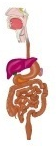
[Shape]
In the living body the shape of the stomach is strongly affected by how full it is and by the posture. Radiographs of the standing person show mainly two types: the J-shaped (hook, or siphon) stomach and the steer-horn stomach. In the J-shaped stomach the body is almost vertical, its lowest point is at about the level of the umbilicus, and the pyloric part lies a little higher and to the right. The steer-horn stomach runs obliquely from upper left to lower right, and the pyloric part is its lowest part. In the cadaver the muscles have lost their tone and the stomach is often swollen like a sac.
[Position]
1. Position in the body
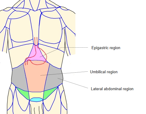
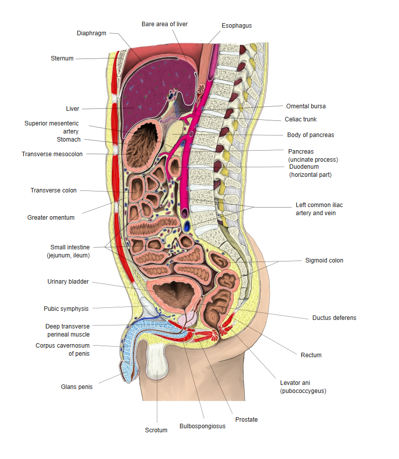
The stomach lies in the upper abdomen (epigastric region and left upper abdomen): most of it (about three quarters) lies in the left lateral part of the upper abdomen and a small part (about one quarter) in its middle part. Its axis runs steeply from upper left and behind to lower right and in front.
2. Position relative to the skeleton
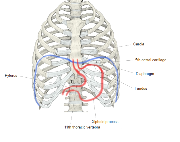
· The cardia lies at the level of the 11th thoracic vertebra, behind the left border of the sternum between the 6th and 7th costal cartilages.
· The pylorus usually lies just to the right of the midline, at about the level of the 1st lumbar vertebra when lying down (lower when standing).
· The lesser curvature corresponds to the left border and lower end of the xiphoid process.
· The right end of the stomach is covered by the liver, in front of which the right costal arch descends.
· The left costal arch crosses the front of the stomach obliquely.
· The fundus rises to about the level of the left 5th costal cartilage.
3. Relations to the surrounding organs
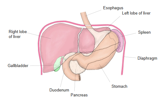
The stomach has important relations to the neighbouring abdominal organs, and several contact areas are distinguished on its surface; they change to some extent with the state of the body and the organs.
· The lesser curvature is covered by the left lobe of the liver; the greater curvature touches the transverse colon.
· On the right, the anterior wall is covered by the quadrate lobe and the left lobe of the liver; on the left, by the costal part of the diaphragm and the anterior abdominal wall.
· The posterior wall touches the lumbar part of the diaphragm and the pancreas.
· The fundus lies to the left of the dome of the diaphragm; behind, it touches the left kidney, the suprarenal gland and the pancreas.
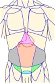 Position in the body
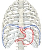 Position relative to
the skeleton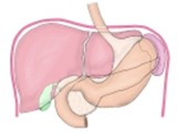 Relations to the organs
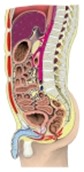 Trunk
(sagittal section)
Parts
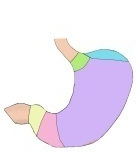
The stomach is divided as follows. See the page of each term for details.
stomach
- Parts of the bag
- Cardia : the region around the upper opening, continuous with the esophagus. Terms: cardia, cardiac orifice, cardiac notch.
- Body : the large middle part of the stomach. Term: gastric canal.
- Fundus : the bulging part above and to the left of the body.
- Pyloric part : the region around the lower opening, continuous with the duodenum. Terms: pylorus, pyloric orifice, pyloric valve.
- Curvatures
- Greater curvature : the lower (greater) border joining the cardia and the pylorus, where the anterior and posterior walls meet.
- Lesser curvature : the upper (lesser) border joining the cardia and the pylorus, where the anterior and posterior walls meet.
- Walls
- Anterior wall : the surface facing forwards and upwards, bounded by the two curvatures.
- Posterior wall : the surface facing backwards and downwards, bounded by the two curvatures.
Structure
stomach wall
- Mucosa
- Epithelium : simple columnar epithelium, renewed rapidly every 3–5 days.
- Lamina propria : loose connective tissue of reticular character, containing the gastric glands and many kinds of cells.
- Muscularis mucosae : two layers of smooth muscle (inner circular, outer longitudinal).
- Submucosa : loose connective tissue made mainly of collagen fibres, with various cells, nerves and vessels.
- Muscle layer
- Outer layer : spreads out radially from the cardia; well developed especially along the greater and lesser curvatures.
- Middle layer : the best developed layer; covers the whole stomach and is especially strong in the pyloric part ⇒ pyloric sphincter, pyloric valve.
- Inner layer : runs obliquely from the cardia and does not reach the pyloric part.
- Serosa : continuous with the peritoneum; covers the anterior and posterior surfaces of the stomach except along the greater and lesser curvatures. Its surface is simple squamous epithelium.
[Mucosa]
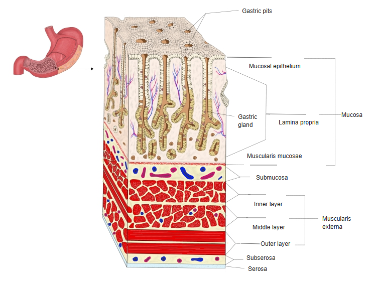
The gastric mucosa has the following three layers.
mucosa
- Epithelium : simple columnar epithelium, renewed rapidly every 3–5 days.
- Lamina propria : loose connective tissue of reticular character, containing the gastric glands and many kinds of cells.
- Muscularis mucosae : two layers of smooth muscle (inner circular, outer longitudinal).
Epithelium
· Simple columnar epithelium of mucus-secreting surface cells; the oval nuclei lie near the base of the cells.
· The upper part of each cell looks pale because its secretory granules have turned into mucus.
· When stimulated, the epithelium secretes more mucus and forms a protective layer on the surface.
· Its power of regeneration is very high, so defects of the gastric mucosa heal fairly easily.
· Mitoses are often seen deep in the gastric pits or at the upper ends of the glands. Undifferentiated epithelial cells that divide in this region (the proliferative zone) differentiate and mature into surface epithelial cells or gland cells.
Lamina propria
· Lymphocytes gather into lymphoid nodules (gastric lymphoid nodules), which are especially numerous in the pyloric part; lymphoid nodules and lymphocytic infiltration are often seen.
Muscularis mucosae
· These smooth muscle fibres are thought to help discharge the secretions of the glands into the lumen of the stomach.
[Submucosa]
· Cells: fat cells, plasma cells, lymphocytes, eosinophils.
· Nerves: the submucosal plexus (Meissner's plexus).
· Vessels: fairly large arteries and veins, and lymph vessels.
[Muscle layer]
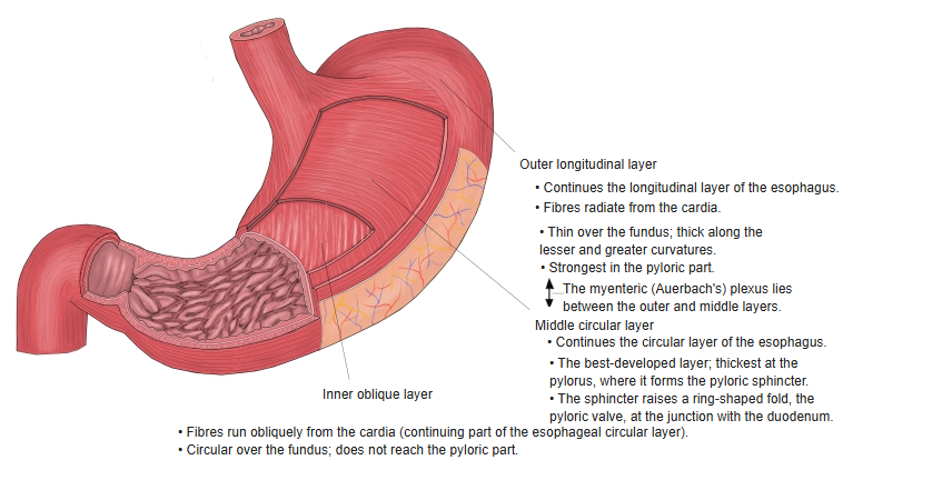
The muscle layer of the stomach is made entirely of smooth muscle and is divided into the following three layers; even the same layer is developed to different degrees in different parts of the stomach.
the stomach wall
- Outer layer : spreads out radially from the cardia; well developed especially along the greater and lesser curvatures.
- Middle layer : the best developed layer; covers the whole stomach and is especially strong in the pyloric part ⇒ pyloric sphincter, pyloric valve.
- Inner layer : runs obliquely from the cardia and does not reach the pyloric part.
Outer layer (longitudinal)
· Continues from the outer longitudinal muscle layer of the esophagus.
· Spreads out radially from the cardia.
· It is weak over the fundus but well developed along the lesser and greater curvatures, where it forms longitudinal bands.
· It is strongest in the pyloric part, especially on its upper and lower surfaces.
· The myenteric plexus (Auerbach's plexus) lies between it and the middle layer.
Middle layer (circular)
· Continues from the inner circular muscle layer of the esophagus and runs around the whole stomach wall.
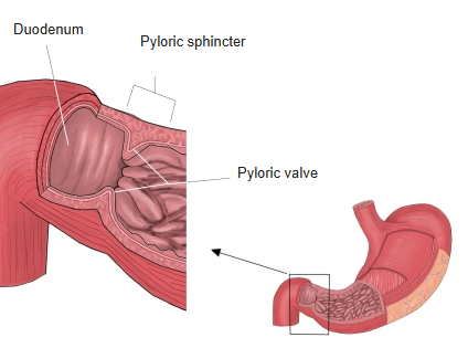
· It is the best developed of the three layers and is especially thick in the pyloric part, where it forms the pyloric sphincter.
· Because of the pyloric sphincter, a ring-shaped fold of mucosa, the pyloric valve, is formed at the junction with the duodenum.
Inner layer (oblique)
· Continues from part of the circular muscle of the esophagus and runs obliquely from the cardia.
· It loops over the fundus but does not reach the pyloric part.
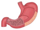 Muscle layers of
the stomach wall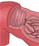 Pyloric sphincter,
pyloric valve
[Serosa]
· The surface of the serosa is covered by simple squamous epithelium (mesothelium); beneath it lies the subserous layer of loose connective tissue rich in nerves and blood vessels.
Gastric glands
- Gastric glands proper : simple tubular glands, branched or unbranched; they form most of the gland body and base, and usually 3–4 open into one gastric pit. = fundic glands. Secrete pepsinogen, hydrochloric acid and mucus.
- Cardiac glands : in the cardia; simple branched tubular mucous glands made of a single cell type.
- Pyloric glands : in the pyloric part; 2–3 open into one gastric pit; mucous, simple branched tubular glands.
The gastric glands are also one of the glands of the digestive system.
digestive system
- Salivary glands : around the oral cavity; divided into the major salivary glands (parotid and others) and the minor salivary glands (palatine, lingual and others).
- Gastric glands : secrete gastric juice (gastric acid, pepsin) from the mucosa; functions: digestion of protein, killing germs, aiding digestion.
- Liver : produces bile (which helps fat digestion); the bile is stored and concentrated in the gallbladder and secreted into the duodenum.
- Pancreas : secretes mainly digestive enzymes as an exocrine gland and hormones as an endocrine gland.
- Duodenal glands : = Brunner's glands; in the submucosa of the duodenum; secrete an alkaline fluid rich in mucus.
- Intestinal glands : in the walls of the small and large intestines; secrete digestive enzymes (peptidases, maltase) and mucus.
[Gastric glands proper (fundic glands)]
· They make up most of the body and base of the glands (in the fundus the glands are sometimes made of chief cells only).
· They are simple tubular glands, branched or unbranched.
· Usually 3–4 glands open into the bottom of one gastric pit.
· Parts: neck, body and base of the gland.
· Secretions: pepsinogen, hydrochloric acid, mucus and hormones. Pepsinogen is the precursor of pepsin; gastric acid or pepsin converts it into pepsin, a protein-digesting enzyme.
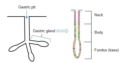
Cells of the gastric glands proper
The gastric glands proper (fundic glands) are made of the following cells.
gastric glands
- Chief cells : form most of the gland body and base; serous cells that secrete pepsinogen.
- Parietal cells : found throughout the gland, most numerous in the body; the largest cells; secrete hydrochloric acid.
- Mucous neck cells : scattered in the neck and the nearby body; the smallest cells; secrete mucus.
- Others : basal-granulated (enteroendocrine) cells, which secrete hormones that regulate digestion.
| 1 | Chief cells | · Make up most of the body and base of the gland; they differentiate from undifferentiated epithelial cells of the proliferative zone, although some chief cells also divide themselves. · Cuboidal to columnar, with indistinct outlines. · Contain fine basophilic granules (containing pepsinogen). · The round nucleus lies at the base of the cell; intercellular secretory canaliculi are present. · Secrete pepsinogen (the precursor of pepsin). · They are serous gland cells. |
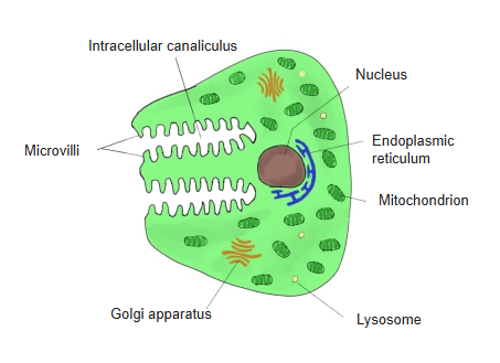
| 2 | Parietal cells (oxyntic cells) | · Found throughout the gland but most numerous in its body. · They differentiate from undifferentiated epithelial cells of the gland neck and live as long as about 3 months. · The largest cells of the fundic glands, 2–3 times as large as the chief cells. · Large, irregularly round or rounded triangular cells with clear outlines. · Have one or two round nuclei in the centre. · Secrete hydrogen ions and chloride ions, which form hydrochloric acid in the intracellular canaliculi (lined with microvilli) and pass into the lumen of the gland. · Contain many large, spherical mitochondria. |
| 3 | Mucous neck cells | · Scattered in the neck of the gland or in the nearby body. · The smallest of the gland cells. · Similar in shape to chief cells, but secrete mucus. · They differentiate from undifferentiated epithelial cells of the proliferative zone and are renewed every 2–8 days. · Rich in neutral or sulfated mucosubstances; their mucus differs in nature from that secreted by the surface mucous cells. · The nucleus lies at the base of the cell and is almost flat. |
| 4 | Others | · Basal-granulated (enteroendocrine) cells, which secrete hormones that regulate digestion. Besides the three kinds above, basal-granulated cells are also fairly numerous; they are found in the cardiac and pyloric glands too. |
[Cardiac glands]
· They are tubular glands in the cardia and are mucous glands closely resembling the pyloric glands.
· The glands are strongly coiled.
· The lumen is wide and lined by a single kind of cell.
· The gland cells are columnar, with round nuclei at the base.
· The part of the cell above the nucleus is filled with mucus.
· Further from the cardia the cardiac glands come to contain chief cells or parietal cells, and are finally replaced by typical fundic glands.
[Pyloric glands]
· They are made of one kind of gland cell, which resembles the mucous neck cells of the gastric glands proper, but with finer and paler granules.
· In the pyloric part the gastric pits are deep, reaching about half or more of the thickness of the mucosa, so the pyloric glands that open into them are short.
· The gland cells closely resemble the mucous neck cells of the fundic glands or the cells of the duodenal glands: they are columnar, with round or oval nuclei at the base.
· The glands are coiled.
· The mucus they secrete is thought to help protect the gastric mucosa; pepsinogen and lysozyme are also found in their secretory granules.
· Very rarely, parietal cells are found in pyloric glands.
Functions
1. Storage of food
Food taken in through the oral cavity is stored for a time in the stomach and then passed on to the small and large intestines for digestion.
2. Digestive juice
The stomach secretes a digestive juice (gastric juice, mainly hydrochloric acid and pepsin) that breaks food down chemically; pepsin in particular breaks down proteins. The strong acidity of the gastric acid kills germs, and the stomach wall protects itself from the acid by secreting mucus.
3. Mixing of food
Peristalsis mixes the food with gastric juice and helps digestion in the intestine.
4. Secretion of hormones
The stomach secretes hormones that regulate digestion (gastrin and others) and promote the activity of the stomach.
| Name | Where secreted | Main actions | |
| 1 | Gastrin | G cells of the pyloric part | · Stimulates the parietal cells and promotes secretion of gastric acid (hydrochloric acid). · Promotes secretion of pepsinogen and helps the digestion of protein. · Promotes the movements of the stomach. · Its secretion increases when food (especially protein) enters the stomach. |
| 2 | Ghrelin | Endocrine cells, mainly in the fundus | · Stimulates appetite, acting on the hypothalamus of the brain to cause hunger. · Promotes secretion of growth hormone (GH). · Activates the movements of the stomach. · Its secretion rises when the stomach is empty and falls after a meal. |
| 3 | Somatostatin | D cells of the pyloric part (also in the pancreas and intestine) | · Inhibits the secretion of gastrin and prevents excessive acid secretion. · Inhibits the secretion of insulin, glucagon and digestive enzymes. · It is secreted when the acidity becomes high, protecting the stomach. |
Vessels and nerves
[Arteries]
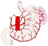
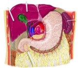
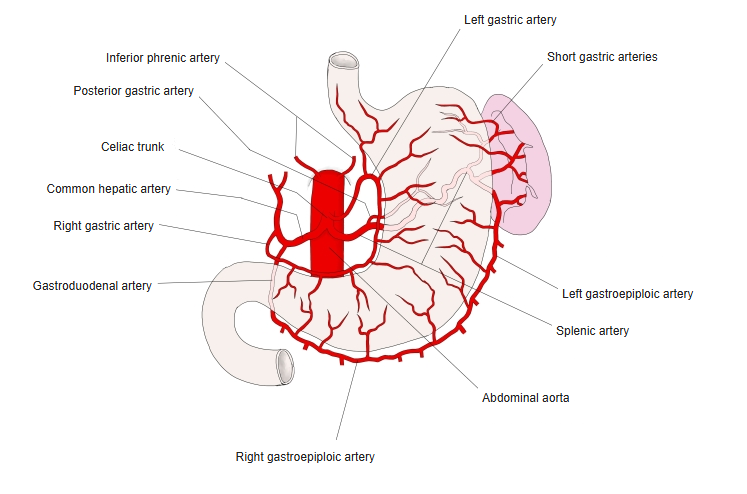
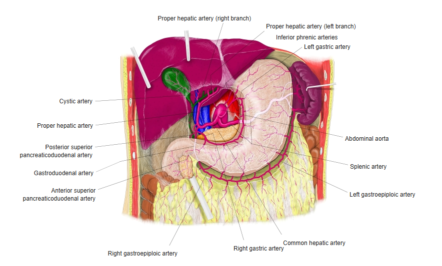
· Cardia and lesser curvature: left gastric artery (a branch of the celiac trunk).
· Lesser curvature and pyloric part: right gastric artery (from the proper or common hepatic artery ← celiac trunk).
· Greater curvature (fundus): short gastric arteries (branches of the splenic artery ← celiac trunk).
· Greater curvature: left gastro-omental (gastroepiploic) artery (a branch of the splenic artery ← celiac trunk).
· Greater curvature (pyloric part): right gastro-omental (gastroepiploic) artery (from the gastroduodenal artery ← common hepatic artery ← celiac trunk).
 Arteries of the stomach
 Arteries around
the stomach
[Veins]
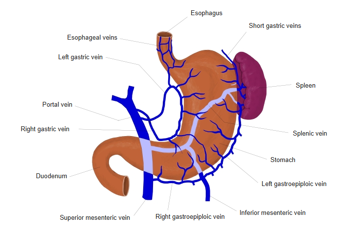
The veins accompany the arteries and, except for part of the cardia, drain into the portal vein: left gastric vein, right gastric vein, short gastric veins, right gastro-omental vein and left gastro-omental vein.
 Veins of the stomach
[Lymphatics]
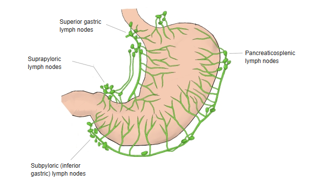
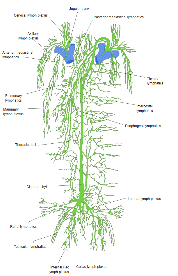
· Lymph passes from the regional lymph nodes through the celiac lymph nodes into the intestinal trunk and then into the cisterna chyli.
· Lymphocytes of the lamina propria gather into lymphoid nodules (gastric lymphoid nodules), which are especially numerous in the pyloric part.
 Lymph nodes around
the stomach Lymph nodes from the thoracic
to the pelvic cavity
[Nerves]
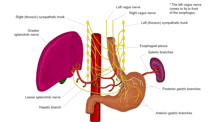
Parasympathetic nerves (vagus nerve, with its gastric branches) and sympathetic nerves (from the greater splanchnic nerve through the celiac ganglia and the celiac plexus, reaching the stomach along its arteries as the gastric plexuses).
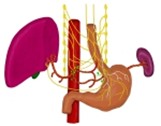 Vagus nerve
(thorax and abdomen)
Related terms
Main anatomical terms related to the stomach, in alphabetical order.
| A | Angular incisure |
| B | Body of stomach |
| C | Cardia, Cardiac glands, Cardiac notch, Cardiac orifice |
| F | Fundus of stomach |
| G | Gastric areas, Gastric canal, Gastric folds, Gastric glands, Gastric pits, Greater curvature |
| L | Lamina propria, Lesser curvature |
| M | Muscularis mucosae |
| P | Pyloric antrum, Pyloric canal, Pyloric glands, Pyloric orifice, Pyloric part, Pyloric sphincter, Pylorus |
| V | Villous folds |
References and sources
- Further reading: Stomach, Wikipedia.