Large intestine (Latin: intestinum crassum)
- Overview
- Structure of the wall
- Mucosa: epithelium (flat parts, crypts), lamina propria, muscularis mucosae, submucosa
- Muscle layer (comparison table, innervation, work)
- Serosa (serosa by part)
- Functions
- Nerves and vessels
- Related terms
- References
Overview
· The large intestine forms faeces, stores them and prepares for their expulsion.
large intestine
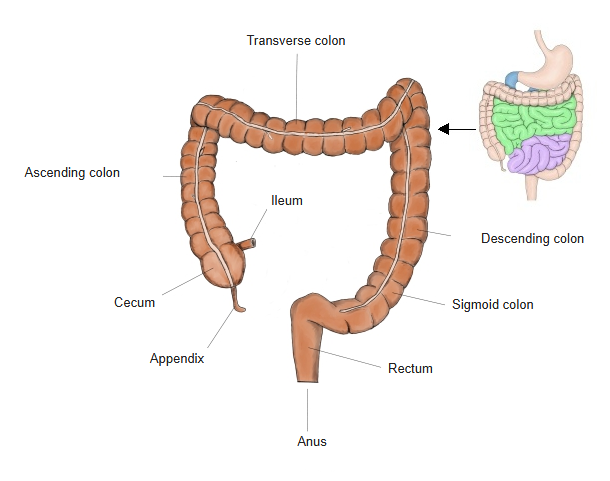
- Cecum : length 6–9 cm; the beginning of the large intestine; temporarily stores the contents from the small intestine.
- Vermiform appendix : a tube about 6–10 cm long; helps to keep the balance of the gut bacteria and takes part in immune function.
- Colon : the main part of the large intestine; functions: absorption of water and electrolytes, storage and transport of faeces. Parts: ascending colon (20–25 cm), transverse colon (40–60 cm), descending colon (20–30 cm), sigmoid colon (30–40 cm).
- Rectum : length 12–15 cm; stores faeces temporarily and passes them to the anal canal for defecation.
- Anal canal : length 3–4 cm; the last part of the digestive tract; faeces are expelled through its end, the anus.
For a detailed description, see the page of each part.
Structure of the wall
large
intestine
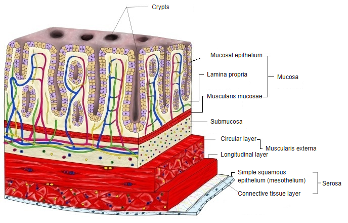
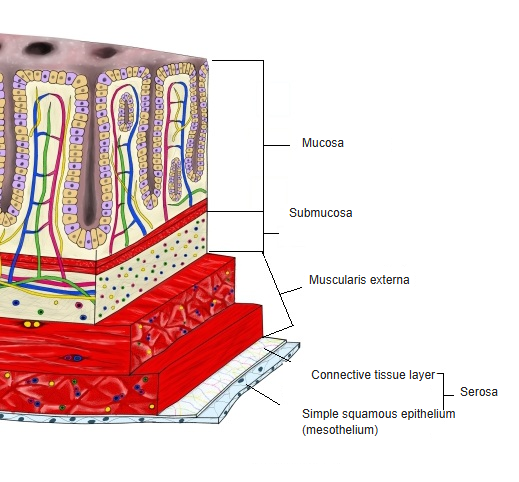
- Mucosa 0.3–0.5
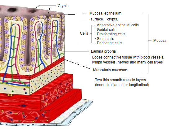
- Mucosal epithelium : flat parts + crypts; greatest feature: no villi; work: recovering water, forming faeces.
- Lamina propria : mainly loose connective tissue; vessels, lymph vessels, nerves and various cells.
- Muscularis mucosae : a thin layer of smooth muscle, 2 layers (inner circular, outer longitudinal).
- → The mucosa does not form circular folds.
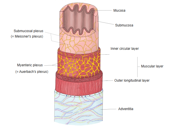
- Submucosa 1–2 : mainly loose connective tissue; vessels, lymph vessels, nerves (submucosal plexus) and various cells.
- Muscle layer 1–1.5
- Inner layer : smooth muscle running round the gut; thicker than the outer longitudinal layer.
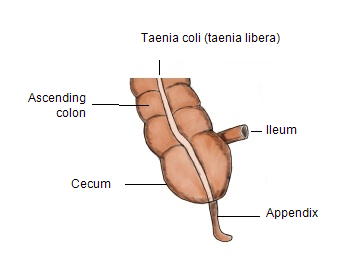
- Outer layer : smooth muscle running lengthwise ⇒ the longitudinal muscle is gathered into the 3 taeniae coli.
- Serosa 0.1–0.2 : simple squamous epithelium (mesothelium) + a thin layer of connective tissue beneath it (vessels, lymph vessels, nerves, a little fat). Parts fused to the posterior abdominal wall (the back of the ascending and descending colon, and others) lack serosa.
* The figures are thicknesses in mm (approximate).
[Mucosa]
of the
mucosa
- Mucosal epithelium
- Flat part : no villi as in the small intestine; also takes part in secretion and absorption.
- Crypts : regularly arranged, straight tubular glands; mucus secretion, absorption, renewal of the epithelium.
- Lamina propria : loose connective tissue, capillaries, lymph vessels and immune cells; lymphoid nodules are seen.
- Muscularis mucosae : a thin layer of smooth muscle, 2 layers: inner (circular) and outer (longitudinal).
■ Mucosal epithelium ■
Main features
1. Specialised for recovering water and forming faeces rather than absorbing nutrients: unlike the small intestine, it does almost no digestion or absorption of nutrients.
2. A thick mucus layer protects the epithelium: there are many goblet cells, which secrete a large amount of mucus (mucin).
3. A structure resistant to physical and chemical stimuli: it is relatively durable and withstands mechanical friction and changes in pH.
⟨Flat part⟩
Cells
| Name | Location | Proportion | Lifespan | Notes | |
|---|---|---|---|---|---|
| 1 | Goblet cells | crypts / surface epithelium (especially many) | 10–20% | 3–7 days | very many (mucus secretion) |
| 2 | Absorptive epithelial cells | whole crypt to surface epithelium | 75–85% | 3–5 days | absorption of water and electrolytes |
⟨Crypts⟩
· Also called the crypts of Lieberkühn (the same in the small intestine).
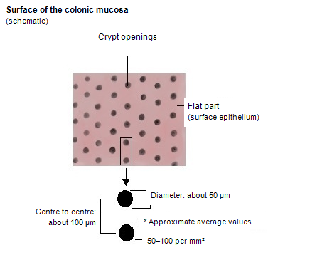
· The crypts are clearly more densely packed than in the small intestine: roughly 50–100 per mm2 in the large intestine and 20–60 per mm2 in the small intestine.
· They are almost straight tubular glands, reaching close to the muscularis mucosae.
Cells
| Name | Location | Proportion | Lifespan | Notes | |
|---|---|---|---|---|---|
| 1 | Absorptive epithelial cells | whole crypt to surface epithelium | 75–85% | 3–5 days | absorption of water and electrolytes |
| 2 | Enteroendocrine cells | mainly the lower crypt | under 1% | several weeks | secrete serotonin |
| 3 | Stem cells | base of the crypt | a few % or less | months to years | lie at the base of the crypt |
| 4 | Proliferating cells | lower to middle crypt | 10–15% | 1–3 days | active cell division |
Enteroendocrine cells: enterochromaffin cells (EC cells, the most numerous), L cells, D cells, N cells, PP cells.
Crypts of the small and large intestines compared
| Item | Small intestine | Large intestine | |
|---|---|---|---|
| 1 | Name of the crypts | crypts of Lieberkühn | crypts of Lieberkühn |
| 2 | Villi | present | absent |
| 3 | Role of the crypts | secretion, supply of cells | secretion, supply of cells (main role) |
| 4 | Length of the crypts | about 200–300 µm | about 300–500 µm |
| 5 | Shape of the crypts | rather short and irregular | long and straight |
| 6 | Diameter of the crypts | about 40–80 µm | about 50–100 µm |
| 7 | Density of the crypts | lower | high |
| 8 | Crypt openings | scattered between the villi | densely packed on the flat mucosa |
| 9 | Main secretion | intestinal juice, antimicrobial factors | mucus (mucin) |
| 10 | Goblet cells | fewer | very many |
| 11 | Part in absorption | indirect | almost none |
| 12 | Stem cells | at the base of the crypt | at the base of the crypt |
| 13 | Paneth cells | present | as a rule absent |
| 14 | Enteroendocrine cells | present | present (somewhat fewer) |
| 15 | Main cells of the surface epithelium | absorptive epithelial cells | absorptive epithelial cells |
| 16 | Points noted in pathology | villous atrophy, crypt hyperplasia | crypt dilatation, branching, atypia |
Flat parts and crypts compared
| Item | Flat part | Crypt | |
|---|---|---|---|
| 1 | Main role | final absorption, forming a barrier | secretion, supply of cells (renewal) |
| 2 | Absorption | yes (main role): water and electrolytes (Na⁺, Cl⁻ and others) | almost none (very slight) |
| 3 | Secretion | almost none | yes (main role), mainly mucus |
| 4 | Mucus secretion | little | very much |
| 5 | Renewal of the epithelium | no | yes (stem cells are present) |
| 6 | Main cells | absorptive epithelial cells | goblet cells, proliferating cells |
| 7 | Proportion of goblet cells | fewer | many |
| 8 | Enteroendocrine cells | almost none | a few |
| 9 | Stem cells | none | at the base of the crypt |
| 10 | Points noted in pathology | surface damage, erosion | crypt dilatation, branching, atypia |
■ Lamina propria ■
· Vessels: a very well-developed capillary network surrounds the crypts (crypts of Lieberkühn).
· Lymph vessels: lymph capillaries often run beside the blood vessels around the bases of the crypts; less conspicuous than in the small intestine.
· Fibroblasts: the main cells of the lamina propria, scattered through the tissue; they secrete collagen and other fibres, hyaluronic acid, proteoglycans and so on.
· Nerves: free nerve endings are present; solitary lymphoid nodules are often seen.
· Immune cells: the lamina propria is a "place of immunity", rich in immune cells. The main ones are lymphocytes (T / B cells), plasma cells (producing IgA: very important), macrophages, dendritic cells and others.
■ Muscularis mucosae ■
· Its functions include fine local movements of the mucosa, helping to empty the contents of the intestinal crypts, and pushing secretions into the lumen.
[Submucosa]
· Vessels: rich in arteries, veins and capillaries; they supply nutrients to the mucosa and collect waste.
· Lymph vessels: receive the flow of lymph from the mucosa.
· Nerves: the submucosal plexus (Meissner's plexus) is found; it takes part in regulating intestinal movement and secretion.
· Cells: fibroblasts secrete collagen fibres, elastic fibres, reticular fibres and ground substance (proteoglycans, hyaluronic acid and others). Immune cells: macrophages, dendritic cells, lymphocytes (T / B cells), neutrophils, eosinophils and others.
Muscle layer
· Inner circular layer: runs round the whole circumference fairly evenly and continuously; the basic structure is the same as in the small intestine. The main player in peristalsis and segmentation.
· Outer longitudinal layer: unlike in the small intestine, it does not cover the whole circumference but forms 3 bands (taeniae coli).
⟨Muscle layers of the digestive tract compared⟩
| Part | Inner circular muscle | Outer longitudinal muscle | Notes | |
|---|---|---|---|---|
| 1 | Esophagus | thick; takes part in peristalsis | upper part voluntary muscle, middle and lower parts smooth muscle | upper esophagus skeletal muscle, lower smooth muscle: mixed |
| 2 | Stomach | thick, with an inner oblique layer added | moderate | besides the inner circular and outer longitudinal layers, the stomach has its own inner oblique layer, allowing mixing (churning) |
| 3 | Small intestine (duodenum, jejunum, ileum) | rather thin; takes part in peristalsis and segmentation | rather thin | the outer longitudinal muscle is even, without bands |
| 4 | Large intestine (colon, rectum) | relatively thick; squeezes the contents | gathered into 3 longitudinal bands | a form peculiar to the large intestine: the banded longitudinal muscle makes the wall bulge (haustra) |
| 5 | Anal part of the rectum | thick, continuous with the anal sphincter | becomes thin | controls defecation together with the anal sphincters |
⟨Innervation⟩
· The enteric nervous system controls the muscle layer.
· Auerbach's (myenteric) plexus: lies between the inner circular and outer longitudinal layers and is in charge of regulating movement.
· It also receives input from the autonomic nervous system (sympathetic and parasympathetic).
⟨Work⟩
1. Peristalsis: the inner circular and outer longitudinal muscles work together to move faeces towards the anus.
2. Segmentation: the inner circular muscle contracts locally → it mixes the contents and promotes the absorption of water.
3. Keeping the shape of the gut: the taeniae coli of the outer longitudinal muscle give the colon its characteristic form.
Serosa
⇒ See serous membrane and mesothelium.
Serosa by part
| Part | Position | Outermost layer | |
|---|---|---|---|
| 1 | Cecum | in the abdominal cavity | serosa |
| 2 | Vermiform appendix | in the abdominal cavity | serosa |
| 3 | Transverse colon | in the abdominal cavity | serosa |
| 4 | Sigmoid colon | in the abdominal cavity | serosa |
| 5 | Ascending colon | retroperitoneal (fixed) | serosa in front only; adventitia behind |
| 6 | Descending colon | retroperitoneal (fixed) | serosa in front only; adventitia behind |
| 7 | Upper rectum | in the pelvis (down to the peritoneal reflection) | partly serosa |
| 8 | Middle and lower rectum | outside the peritoneum | adventitia |
Functions
The main work of each part is listed below.
| Part | Main work | Notes | |
|---|---|---|---|
| 1 | Cecum | receiving the contents; site of fermentation | receives the contents from the small intestine and makes an environment where gut bacteria work easily |
| 2 | Vermiform appendix | immunity; store of gut bacteria | takes part in immunity and acts as a "store" of beneficial bacteria |
| 3 | Ascending colon | absorption of water and electrolytes | an important site of absorption where the faeces begin to harden |
| 4 | Transverse colon | absorption + transport | continues absorbing while sending the faeces to the left |
| 5 | Descending colon | concentrating and storing faeces | stores the fairly hard faeces for a time |
| 6 | Sigmoid colon | regulating defecation; final storage | a store that adjusts the timing of defecation |
| 7 | Rectum | triggering the defecation reflex | senses the faeces and produces the urge to defecate |
| 8 | Anal canal | controlling expulsion | controls the passage of faeces with the sphincters |
Nerves and vessels
· Derived from the midgut: cecum, appendix, ascending colon, transverse colon (proximal two-thirds).
· Derived from the hindgut: transverse colon (distal one-third), descending colon, sigmoid colon, upper rectum (about two-thirds).
* The lower rectum is derived from the cloaca, and the lower anal canal below it from the ectoderm.
· Arteries: the midgut part is supplied by the superior mesenteric artery, the hindgut part by the inferior mesenteric artery; the veins follow the same pattern (superior and inferior mesenteric veins).
[Nerves]
· Enteric nervous system (ENS): the gut's own nervous system; it takes part in peristalsis, secretion and so on without going through the central nervous system.
nerve
plexuses
- Submucosal plexus : = Meissner's plexus; the nerve plexus in the submucosa, outside the mucosa.
- Intermuscular (myenteric) plexus : = Auerbach's plexus; the plexus between the inner circular and outer longitudinal layers.
- Mucosal plexus : the plexus in the mucosa, especially near the muscularis mucosae. Standard anatomy textbooks often do not include it.
- → These are found along the whole digestive tract.
Sympathetic: thoracic and lumbar splanchnic nerves (lesser splanchnic nerve and others); work: inhibits movement and secretion, constricts blood vessels.
Parasympathetic: vagus nerve (proximal two-thirds of the transverse colon and above) and pelvic splanchnic nerves (beyond); work: promotes peristalsis and secretion.
* Near the middle of the transverse colon the parasympathetic supply changes from the vagus nerve to the pelvic splanchnic nerves.
of the large
intestine
- Enteric nervous system : the main innervation of the gut; exists fairly independently of the central nervous system.
- Myenteric plexus : between the inner circular and outer longitudinal layers = Auerbach's plexus
- Submucosal plexus : in the submucosa = Meissner's plexus
- Autonomic nervous system : mainly synapses with the enteric nervous system; part of it acts directly on the vessels and sphincters.
- Sympathetic [thoracic and lumbar splanchnic nerves] : inhibits peristalsis and secretion; contracts the vessels and sphincters.
- Parasympathetic [vagus nerve (proximal 2/3), pelvic splanchnic nerves (distal 1/3)] : promotes peristalsis, secretion and blood flow.
- Sensory nerves : belong to the central nervous system; they run with the autonomic nerves to all layers of the wall. Pain is felt not evenly but mainly from the muscle layer and serosa.
- With the sympathetic: visceral afferent fibres carry pain, ischaemia and inflammation to the centre.
- With the parasympathetic: visceral afferent fibres carry physiological sensations and reflex input to the centre.
Related terms
Main anatomical terms related to the large intestine, in alphabetical order.
| C | Cecum, Colon, Crypts of Lieberkühn, Circular folds |
| E | Enteric nervous system, Enterocyte, Enteroendocrine cell |
| G | Goblet cell |
| H | Haustra of the colon |
| M | Meissner's plexus, Mesothelium, Myenteric plexus |
| R | Rectum |
| S | Serous membrane, Submucosa, Submucosal plexus |
| T | Taeniae coli |
| V | Vermiform appendix, Villi |
References and sources
- Further reading: Large intestine, Wikipedia.