Foramina of the cranial base (Latin: foramina basis cranii)

Also called Neurovascular pathways through the base of the skull

The cranial base is pierced by openings of various sizes through which vessels and nerves, including most of the cranial nerves, pass between the cranial cavity and the face, orbit and neck. Seen from inside, they lie in the floors of the anterior, middle and posterior cranial fossae; some, such as the palatine foramina and the incisive foramen, are seen only on the external surface. The table below lists each opening, the bone in which it lies, and the nerves, arteries and veins that pass through it. Each opening is linked to its own page.

Textbooks differ on some of the smaller vessels, so the names in the table may not match every source or every page of the single openings; the main differences are given in the notes below the table.

Openings (A-Z): Carotid canal, Condylar canal, Cribriform plate, Foramen lacerum, Foramen magnum, Foramen ovale, Foramen rotundum, Foramen spinosum, Greater palatine foramen, Hiatus for greater petrosal nerve, Hiatus for lesser petrosal nerve, Hypoglossal canal, Incisive foramen, Internal acoustic opening, Jugular foramen, Lesser palatine foramina, Mastoid foramen, Optic canal, Petrotympanic fissure, Stylomastoid foramen, Superior orbital fissure.

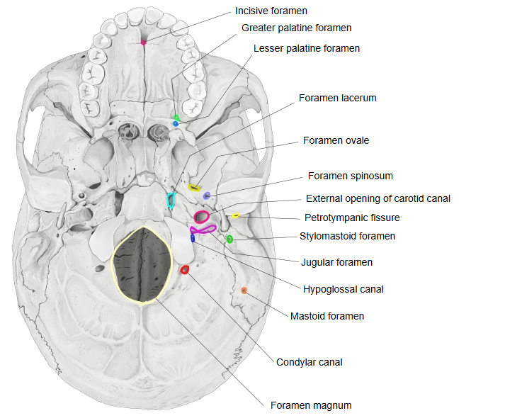
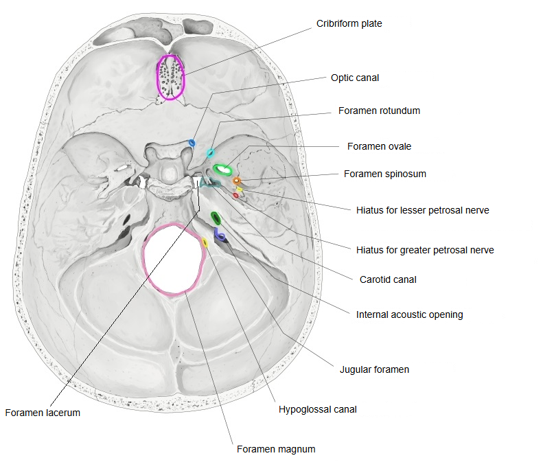
⇒ Illustrations: see the two figures at the end of the table notes.

1Cribriform plate   Bone: ethmoid bone
Nerves: olfactory nerves, anterior ethmoidal nerve
Arteries: anterior ethmoidal artery, posterior ethmoidal artery
2Optic canal   Bone: sphenoid bone (lesser wing)
Nerves: optic nerve
Arteries: ophthalmic artery
3Superior orbital fissure   Bone: sphenoid bone (between the greater wing and the lesser wing)
Nerves: oculomotor nerve, trochlear nerve, abducens nerve, the branches of the ophthalmic nerve, sympathetic nerve fibres from the cavernous plexus around the internal carotid artery
Veins: superior ophthalmic vein
4Foramen rotundum   Bone: sphenoid bone (greater wing)
Nerves: maxillary nerve
5Foramen ovale   Bone: sphenoid bone (greater wing)
Nerves: mandibular nerve, with the motor root of the trigeminal nerve; lesser petrosal nerve (often)
Arteries: accessory meningeal branch
Veins: emissary veins (venous plexus of foramen ovale), some middle meningeal veins
6Foramen spinosum   Bone: sphenoid bone (greater wing)
Nerves: meningeal branch of the mandibular nerve
Arteries: middle meningeal artery (branch of the maxillary artery)
Veins: middle meningeal veins
7Carotid canal (external and internal openings)   Bone: temporal bone (petrous part)
Nerves: internal carotid plexus
Arteries: internal carotid artery
Veins: internal carotid venous plexus
8Hiatus for lesser petrosal nerve   Bone: temporal bone (petrous part)
Nerves: lesser petrosal nerve
Arteries: superior tympanic artery (note 1)
9Hiatus for greater petrosal nerve   Bone: temporal bone (petrous part)
Nerves: greater petrosal nerve
Arteries: petrosal branch of the middle meningeal artery
10Internal acoustic opening   Bone: temporal bone (petrous part)
Nerves: facial nerve (with the intermediate nerve), vestibulocochlear nerve
Arteries: labyrinthine artery
Veins: labyrinthine veins
11Jugular foramen   Bones: temporal bone, occipital bone
Nerves: glossopharyngeal nerve, vagus nerve, accessory nerve
Arteries: posterior meningeal artery (from the ascending pharyngeal artery)
Veins: inferior petrosal sinus; the sigmoid sinus continuing as the internal jugular vein
12Foramen magnum   Bone: occipital bone
Nervous system: lower end of the medulla oblongata, continuing into the spinal cord, with its meninges; spinal roots of the accessory nerves (note 2)
Arteries: vertebral arteries, anterior and posterior spinal arteries
Veins: anterior and posterior spinal veins; the link between the internal vertebral venous plexus and the basilar plexus
13Condylar canal   Bone: occipital bone
Veins: condylar emissary vein
14Hypoglossal canal   Bone: occipital bone
Nerves: hypoglossal nerve
Arteries: a meningeal branch of the ascending pharyngeal artery
Veins: venous plexus of the hypoglossal canal
15Mastoid foramen   Bone: temporal bone
Arteries: meningeal branch of the occipital artery
Veins: mastoid emissary vein
16Stylomastoid foramen   Bone: temporal bone
Nerves: facial nerve
Arteries: stylomastoid artery
Veins: stylomastoid vein
17Petrotympanic fissure   Bone: temporal bone (between the petrous part and the tympanic part)
Nerves: chorda tympani
Arteries: anterior tympanic artery
Veins: tympanic veins
18Foramen lacerum (note 3)   Bones: sphenoid bone, temporal bone (apex of the petrous part) and occipital bone (basilar part)
Veins: emissary vein of foramen lacerum (note 4)
19Lesser palatine foramina   Bone: palatine bone
Nerves: lesser palatine nerves (see palatine nerves)
Arteries: lesser palatine arteries
Veins: lesser palatine veins? (note 5)
20Greater palatine foramen   Bone: palatine bone (with the maxilla)
Nerves: greater palatine nerve
Arteries: greater palatine artery
Veins: greater palatine vein
21Incisive foramen   Bone: maxilla
Nerves: nasopalatine nerves
Arteries: nasopalatine artery (posterior septal branch of the sphenopalatine artery), joined by the end of the greater palatine artery

Note 1: Sources differ on the route of the superior tympanic artery, a branch of the middle meningeal artery. Some textbooks describe it entering the temporal bone with the lesser petrosal nerve, others through the hiatus for the greater petrosal nerve.

Note 2: The first cervical nerve does not pass through the foramen magnum; it leaves the vertebral canal between the occipital bone and the atlas as the suboccipital nerve.

Note 3: In life the foramen lacerum is closed by cartilage, and almost nothing passes all the way through it: only small emissary veins and a meningeal branch of the ascending pharyngeal artery. The internal carotid artery crosses its upper part on its way from the carotid canal to the cavernous sinus. Over the cartilage, the greater petrosal nerve and the deep petrosal nerve join to form the nerve of the pterygoid canal, which runs forward with the artery of the pterygoid canal into the pterygoid canal.

Some textbooks and atlases therefore list the greater and deep petrosal nerves, or the nerve and artery of the pterygoid canal, as passing through the foramen lacerum, while others list nothing; sources differ.

Note 4: The emissary vein of foramen lacerum is a small vein that passes through the foramen lacerum and joins the cavernous sinus with the pterygoid plexus.

Note 5: Some sources name lesser palatine veins running through the lesser palatine foramina with the arteries, but most textbooks do not describe them.
  • The base of the skull from below, with each opening outlined in colour and named.External cranial base
  • The floor of the cranial cavity from above, with each opening outlined in colour and named.Internal cranial base

References and sources

  1. Further reading: List of foramina of the human body, Wikipedia.
  2. Further reading: Base of skull, Wikipedia.
  3. Federative International Programme for Anatomical Terminology (FIPAT). Terminologia Anatomica, 2nd ed. (TA2), 2019.